


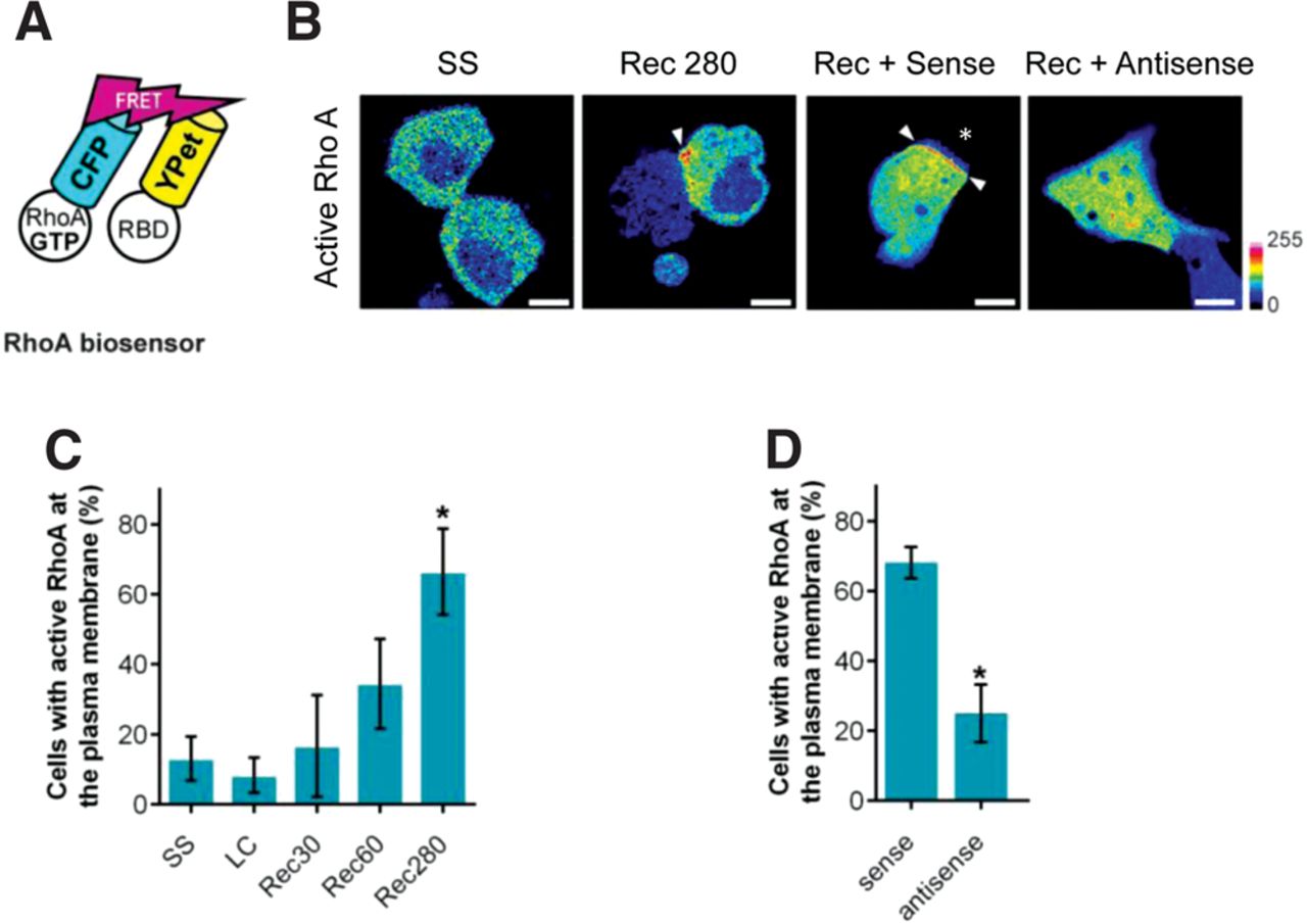
Active-RhoA accumulates and persists at cell contact sites during the contact expansion stage of epithelial AJ assembly. An intermolecular FRET-based RhoA biosensor was expressed in MDCK cells to visualize the subcellular localization of active-RhoA following epithelial cell contact. (A) The intermolecular FRET-based RhoA biosensor consists of RhoA tagged to CFP (donor) and the amino acids 7–89 of Rhotekin fused to Ypet (acceptor). The activation-dependent binding of the donor and acceptor was visualized by sensitized-emission FRET microscopy. (B) RhoA activity is targeted to contact site-localized plasma membrane during cell junction formation. Under steady state (SS) conditions in confluent epithelial cell monolayers, active-RhoA is uniformly distributed throughout the cytoplasm. During junction assembly following incubation in Ca2+ recovery media (Rec 280), active-RhoA is targeted to regions of the plasma membrane adjacent to sites of cell contact. MDCK cells expressing the RhoA biosensor were treated as indicated, and RhoA activity was imaged by sensitized-emission microscopy. Sense oligonucleotides illustrated contact-localized active-RhoA at the cell contact (Rec + Sense). Antisense oligomers targeting β-actin zipcode mRNA prevent recruitment of active RhoA to the plasma membrane (Rec + Antisense). MDCK cells expressing the RhoA biosensor were treated as indicated, and RhoA activity was imaged between 1 and 2 h following the return to the Ca2+ recovery media. The images were pseudocolored according to the scale to the right of the figure. Scale bars, 5 µm. Arrowheads indicate regions of active RhoA at the plasma membrane. (C) Quantification of RhoA activity distribution during cell junction formation. (*) P < 0.05 vs. LC. Error bars show means ± SEM based on a minimum of 37 cells. (D) The distribution of RhoA activity was quantified in cells treated as indicated. (*) P < 0.05 vs. WT sense. Error bars show means ± SEM based on a minimum of 17 cells.




