


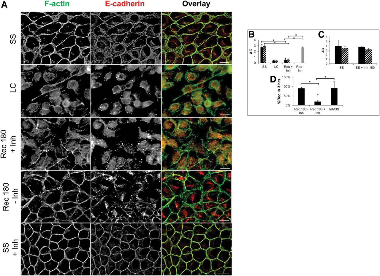
De novo protein synthesis is required to assemble but not maintain epithelial AJ complexes. (A) De-convolved epifluorescence images of a single plane of MDCK cells at steady state (SS), cells treated with low Ca2+ media for 1 h (LC), or cells after 3 h in Ca2+-containing recovery media without (Rec 180-Inh) or with (Rec 180-Inh) cycloheximide during Ca2+ switch experiments. F-actin (green) is stained with Alexa488-phalloidin, and E-cadherin is stained by immunofluorescence (red), with numerous AJ complexes (yellow) observed at cell contact sites. Cycloheximide was also applied to monolayers at steady state (SS + Inh) for 3 h. (Boxed panel) Graphs represent the asymmetry coefficient (AC) and percent recovery (%Rec) during MDCK Ca2+switch experiments. (B) AC during Ca2+ switch experiments without translation inhibitors (gray bar) or with translation inhibitors, cycloheximide (black bars), or puromycin (striped bars). (C) AC when translation inhibitors cycloheximide (black bars) or puromycin (striped bars) are added to steady state MDCK monolayers without undergoing a Ca2+ switch. (D) Summary of the %Rec for the Ca2+ switch with and without translation inhibitors, including the direct application of translation inhibitors to the steady-state monolayers. Scale bars, 20 μm. Error bars represent mean ± SEM based on five cells from three independent experiments. (*) P < 0.05.




