


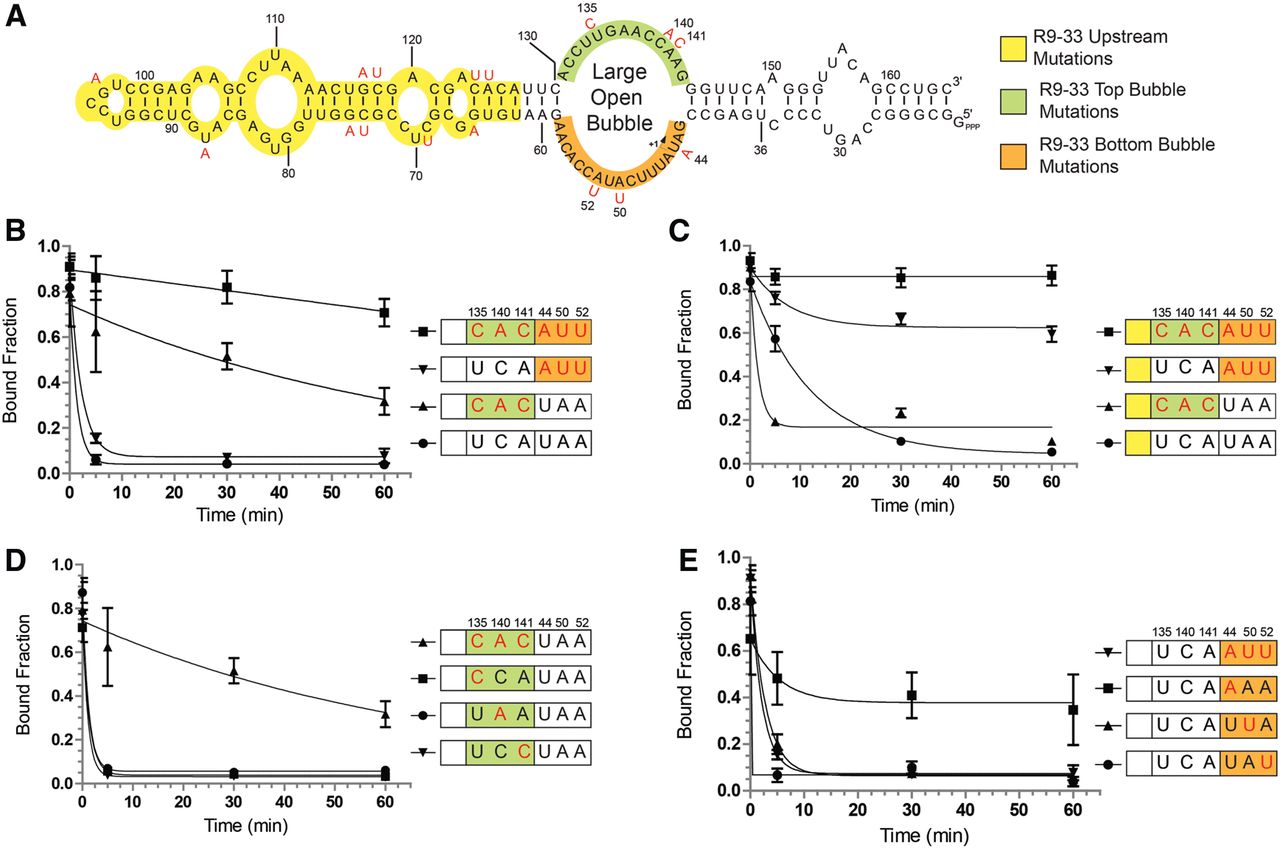
Substitutions around the large open bubble (LOB) of R9-33 act in a highly synergistic manner and are responsible for blocking release from Eσ70. (A) T1 RNA sequence is noted in black while substitutions found in R9-33 are in red letters. The molecule was divided into three distinct zones: a yellow upstream region that contains 10 mutations; a green top strand in the LOB region contains three substitutions at positions 135, 140, and 141; an orange bottom strand in the LOB contains three substitutions at positions 44, 50, and 52. (B) Release kinetics for T1 RNA and three other constructs with triple mutations (in red letters). (C) Release kinetics for R9-33 constructs with substitutions that were sequentially removed according to the color code described in panel A. (D) Release kinetics for constructs with point mutations (in red letters) in the top strand of the 6S RNA LOB. (E) Release kinetics for constructs with point mutations (in red letters) in the bottom strand of the 6S RNA LOB. Error bars correspond to standard deviations about the mean of at least three independent experiments.




