


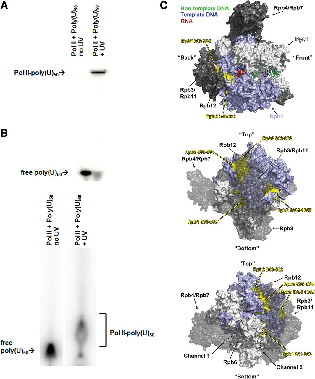
Binding of poly(U) RNA to Pol II subunits. (A) Purified Pol II was bound to poly(U)50 RNA, either with or without UV crosslinking. Complexes were denatured with formamide and subjected to denaturing polyacrylamide gel electrophoresis to isolate subunits that become attached to the poly(U). The large shifted band in the presence of Pol II is usually a doublet, consistent with both Rpb1 and Rpb2 shifts. Binding reactions using Pol II and 32P-labeled poly(U) +/− UV crossinking, shown, were run in parallel with identical unlabeled samples for excising shifted bands and analysis (Table 1). (B) Pol II was crosslinked to poly(U)50 as in A, but before denaturation and electrophoretic separation, the complex was exhaustively digested with trypsin. Peptides reproducibly associated with the indicated shifted region were determined (Table 2). (C) Views of the Pol II holoenzyme (PDB 3po2, 51), with peptides from the shifted poly(U) band in C shown in yellow. PDB entry 3po2 contains 12-subunit Pol II containing a nucleic acid scaffold comprising a template DNA strand (blue), a partially visible nontemplate strand (green), and a product RNA (red), illustrating RNA in an RNA-DNA hybrid with template DNA with a number of bases present in a long backtrack (not visible in the views shown). (Top) 12-subunit Pol II holoenzyme, illustrating the “crab claw” shape of Pol II with the large subunits (Rpb1 in white, Rpb2 in light blue) comprising one claw each, with template DNA and RNA-DNA hybrid bound between them. Other subunits are in gray, with Rpb4/Rpb7 forming a stalk to one side of Pol II. (Middle) “Back view” of Pol II from behind, with Rpb3-Rpb12 rendered as partially transparent. (Bottom) “Back view” as above but tilted toward Rpb4/Rpb7 to illustrate positions of possible RNA channels 1 and 2. Channel 1 is experimentally supported as the RNA exit channel, whereas Channel 2 is electrostatically favorable for RNA interaction.




