


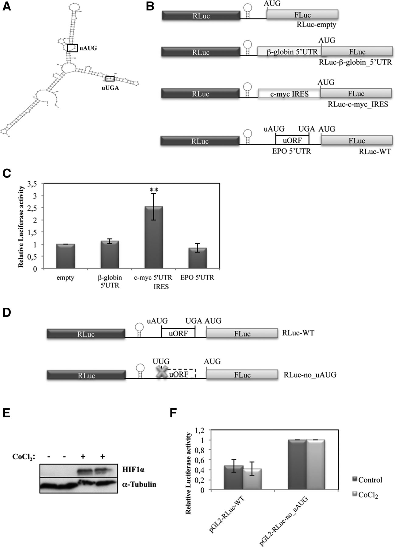
EPO translational derepression in response to hypoxia in HeLa cells is not mediated by an IRES. (A) Representation of the secondary structure of the 5′-leader sequence of the human EPO transcript as predicted by the Mfold webserver. (B) Schematic of the dicistronic luciferase vectors. The 5′ UTR sequence of the human EPO transcript (EPO 5′ UTR) to be tested for IRES activity, as well as the 5′ UTR sequence of the human β-globin transcript (β-globin 5′ UTR), or the c-myc IRES sequence previously described by Stoneley et al. (1998), was inserted between the Renilla (RLuc) and firefly (FLuc) luciferase cistrons, downstream from a hairpin structure (represented by a stem–loop) in the multiple cloning site spacer of the RLuc-empty vector to create the RLuc-WT, RLuc-β-globin_5′ UTR, and the RLuc-c-myc_IRES constructs, respectively. (C) HeLa cells were transiently transfected with each one of the constructs described in B and with a plasmid encoding RLuc (pRL-TK) and analyzed as described in the legend to Figure 2B. (D) Schematic of the dicistronic reporter constructs used to test if the EPO 5′-leader sequence contains an IRES activated during hypoxia. RLuc-WT contains the human EPO 5′-leader sequence with the intact uORF, as defined in A, and the RLuc-no_uAUG contains the EPO 5′-leader sequence with a disrupted uORF due to the uAUG→UUG mutation (represented by a cross). HeLa cells were transfected with these constructs. Six hours later, cells were untreated or treated with 200 µM CoCl2 for 24 h. (E) Representative Western blot analysis of HeLa cell extracts untreated (−) or treated (+) with CoCl2. Immunoblotting was performed using a human HIF1α–specific antibody to control the stress conditions and a human α-tubulin–specific antibody to control for variations in protein loading. (F) Relative luciferase activity was quantified as described in the legend to Figure 2B.




