


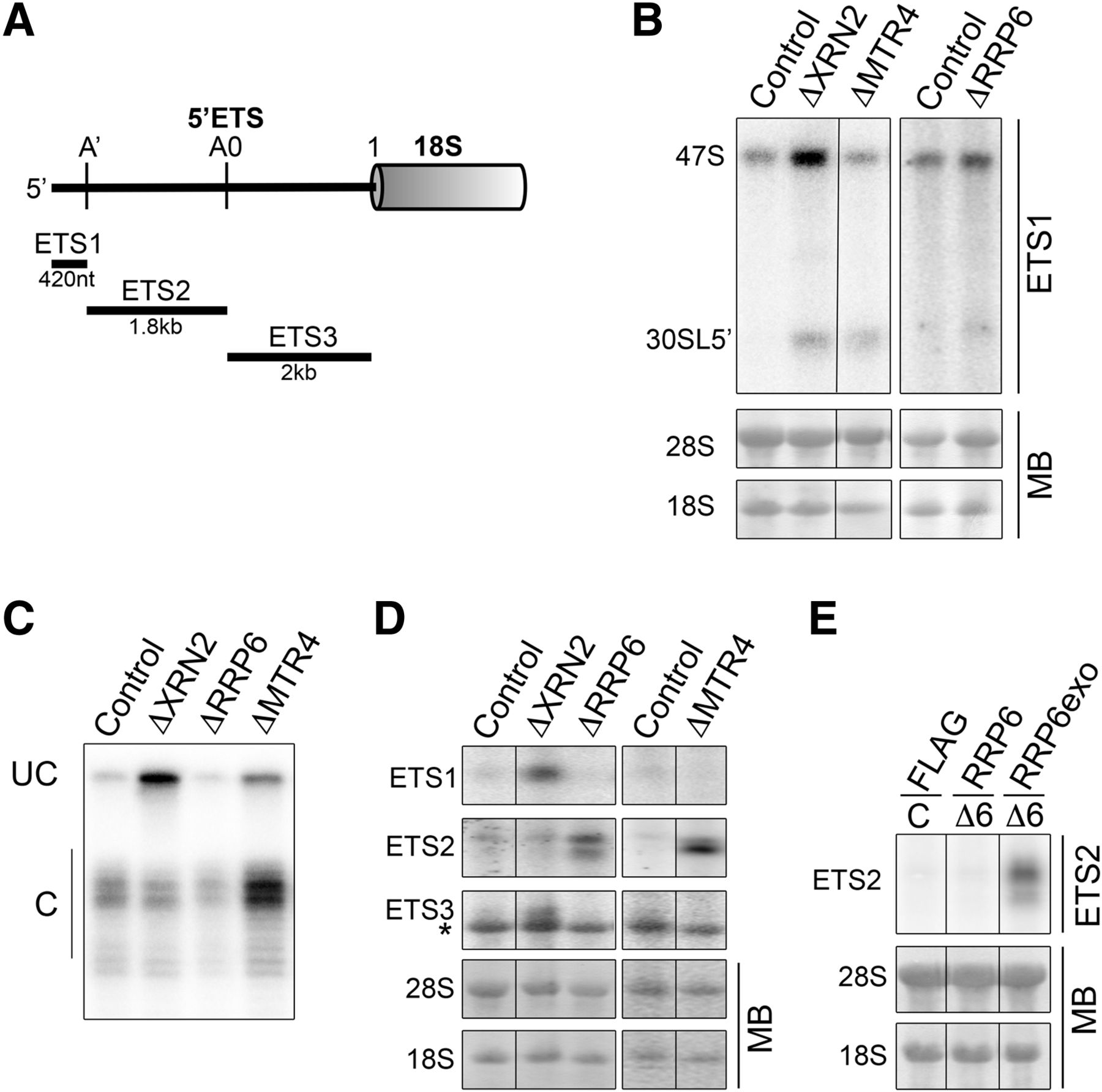
XRN2, RRP6, and MTR4 function in A′ cleavage and turnover of 5′ETS fragments. (A) Schematic outline of the metazoan 5′ETS showing the cleavage sites and fragments generated. (B) RNA from HeLa cells depleted of XRN2, RRP6, or MTR4 was separated by agarose-glyoxal gel electrophoresis and analyzed by Northern blotting using an ETS1 probe. Mature rRNAs were visualized using methylene blue staining. (C) RNA from siRNA-treated cells was analyzed by S1 nuclease mapping using a probe hybridizing across the A′ cleavage site. The resulting DNA fragments ([UC] uncleaved, [C] cleaved) were separated by denaturing polyacrylamide gel electrophoresis and visualized using a phosphorimager. Note that, relative to the control, the ▵RRP6 lane is underloaded. (D) HeLa cells were transfected with control siRNAs or those targeting XRN2, MTR4, or RRP6, and 60 h later, RNA was harvested. This was analyzed by agarose-glyoxal gel electrophoresis followed by Northern blotting using probes hybridizing between the 5′-A′ (ETS1), A′-A0 (ETS2), and A0-site 1 (ETS3) cleavage sites. Note that the ETS3 probe cross-reacts with 18S—indicated by an asterisk (*). (E) HEK293 cells stably transfected with plasmids enabling expression of the FLAG-tag alone, FLAG-RRP6, or an exonucleolytically inactive form of RRP6 (RRP6exo) were transfected with control siRNAs (C) or those against RRP6 (Δ6). RNA was analyzed by Northern blotting using an ETS2 probe.




