


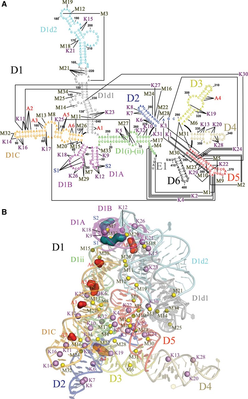
FIGURE 1.
Site-bound ions in O. iheyensis group II intron. (A) Secondary structure map of the crystallization construct adapted from Pyle (2010) to indicate the site-bound ions described in this work. D6 is included for completeness, but it was not present in the crystallization construct and its structure is currently undetermined. E1 indicates the four nucleotides of the 5′-exon. (B) Overall crystal structure in which all site-bound ions are represented as spheres. M sites are yellow, K sites violet, S sites blue, and A sites red. Each intron subdomain is depicted in a different color.




