


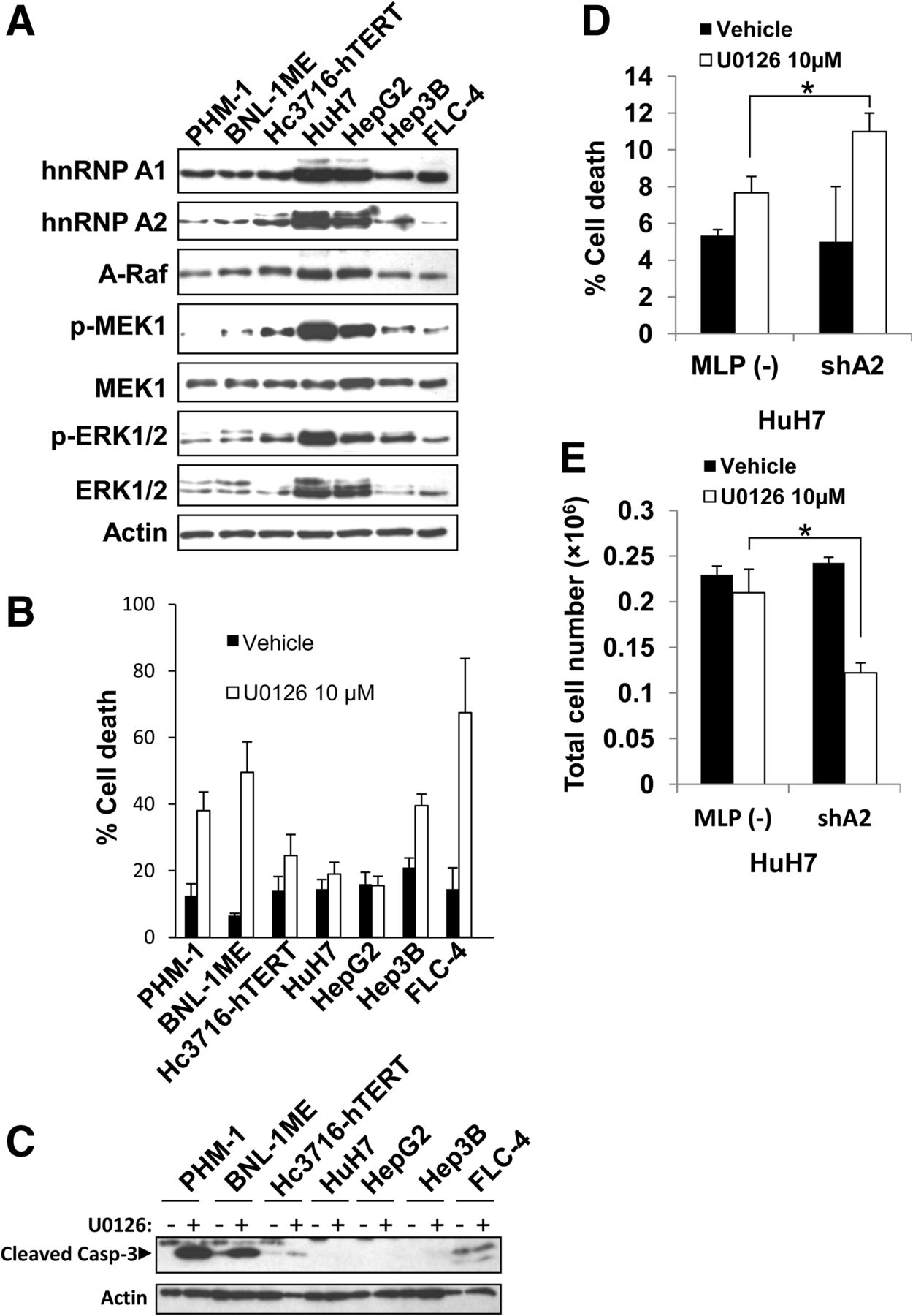
Sensitivity of HCC cells to MEK inhibitor–induced apoptosis correlates with hnRNP A1 and A2 levels. (A) Cells were lysed, and protein levels of hnRNP A2/B1, hnRNP A1/A1b, A-Raf FL and total and phosphorylated MEK1/2 and ERK1/2 were examined using Western blot. β-Actin was used as loading control. (B) HCC cell lines were treated either with vehicle or MEK inhibitor U0126 (10 µM) for 24 h and subjected to trypan blue exclusion assay. (C) Cells described in A and B were analyzed by Western blot for cleaved caspase-3, a marker for apoptosis. β-Actin served as loading control. (D) HuH7 cells transduced with the indicated retroviruses were seeded (120 × 104 cells/well in a six-well plate in triplicates) and treated with either vehicle or MEK inhibitor U0126 (10 µM) for 48 h and subjected to trypan blue exclusion assay. (*) Increased apoptosis after U0126 treatment in hnRNP A2 knockdown compared with MLP. P = 0.053. (E) Cells described in D were counted (total cell number) by a BioRad cell counter. (*) Reduced cell number after U0126 treatment in hnRNP A2 knockdown compared with MLP. P = 0.033. Experiments described in D and E were repeated at least twice.




