


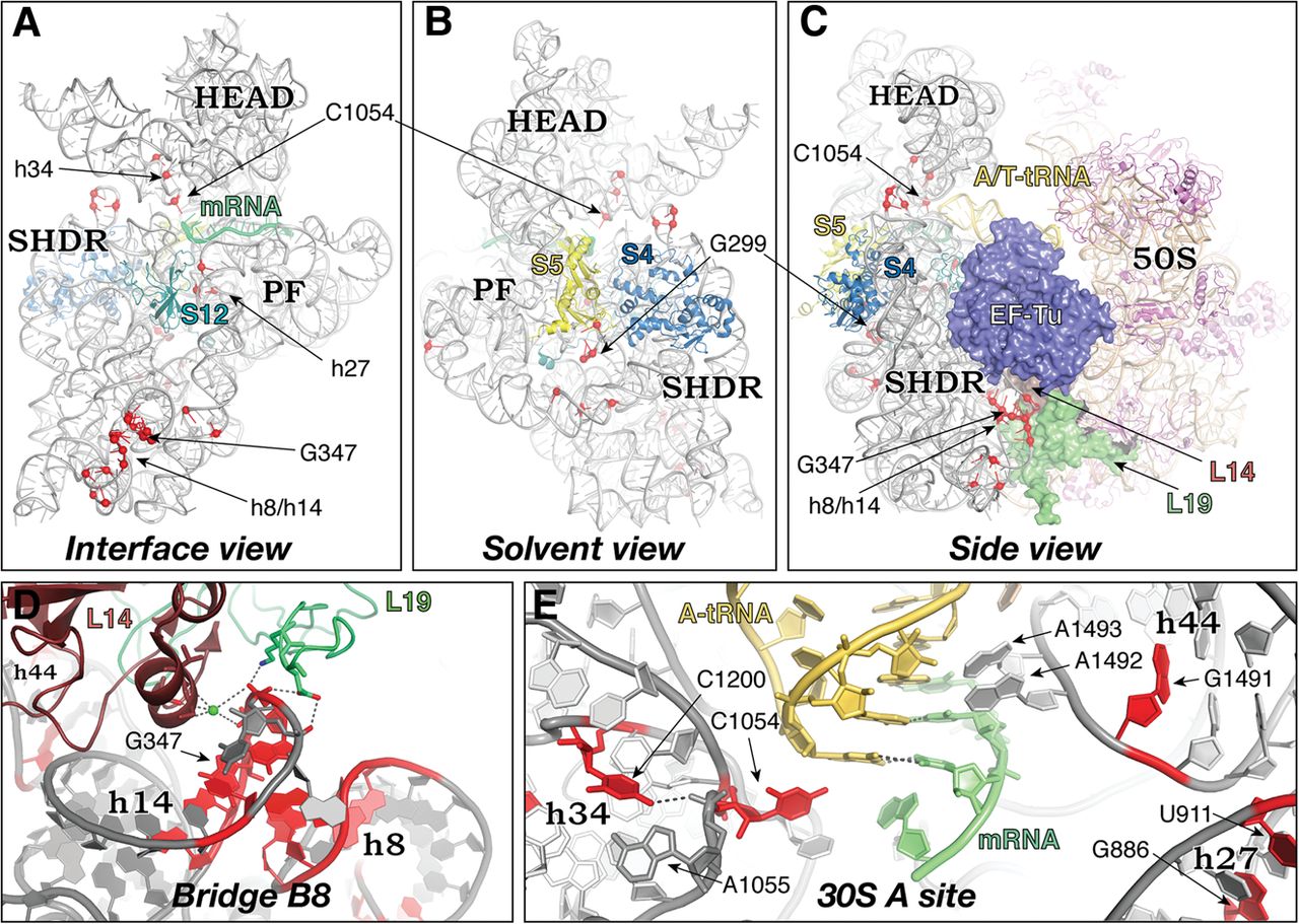
Locations of ram mutations in 16S rRNA. Tertiary structure of the 16S rRNA, viewed from the subunit interface (A) and solvent (B) perspectives, and in the context of the 70S ribosome with bound ternary complex (C). Red spheres indicate positions of ram mutations. Small subunit proteins other than S4, S5, and S12 have been computationally removed for clarity. (SHDR) Shoulder domain, (PF) platform domain. (D) Zoomed-in view of intersubunit bridge B8. Hydrogen bonds that are lost upon bridge disruption are shown as dashed lines. (E) Zoomed-in view of the 30S A site showing the interactions of various 16S rRNA residues with A-tRNA and mRNA. For clarity, h18 and S12 are omitted from the foreground of this view. (Gray) 16S rRNA, (tan) 23S/5S rRNA, (magenta) unlabeled 50S proteins, other features as indicated. Figure based on PDB files 2WRN, 2WRO, 2WDG, and 2WDI.




