


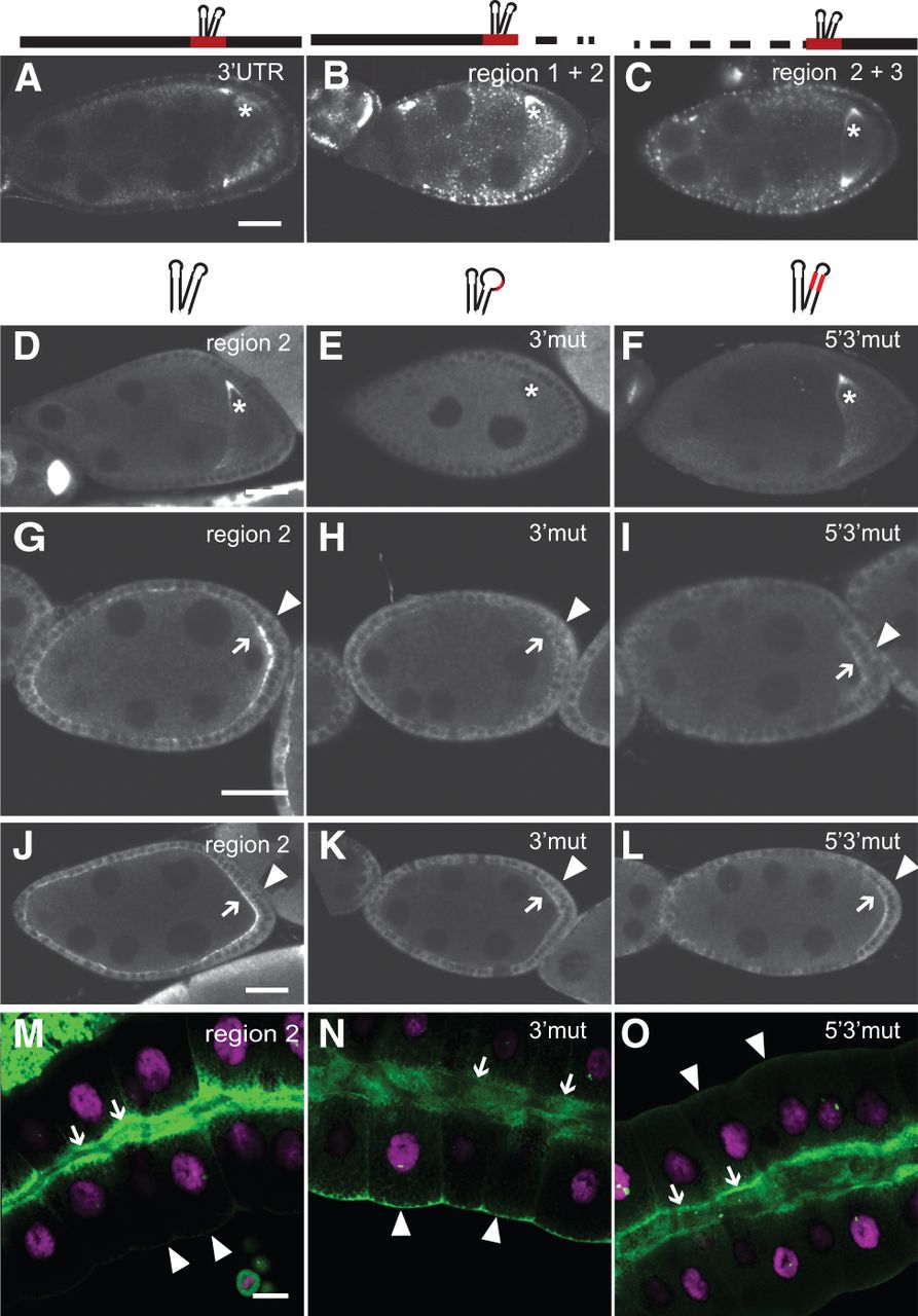
The oocyte entry signal mediates anterior localization in stage 8 oocytes and apical localization in polarized epithelia. (A–O) Transgenic UAS-oskar 3′ UTR reporters fused to the egfp open reading frame were expressed under the control of the mat-α4-tub-Gal4 in the germline (A–F), GR1-Gal4 in follicular epithelial cells (G–L), or forkhead-Gal4 in salivary glands (M–O). Reporter RNAs were detected by fluorescent in situ hybridization using an egfp-antisense probe (RNA shown in green in M–O). For orientation, the salivary glands (M–O) were counter-stained with DAPI to reveal DNA (shown in magenta). A star indicates the position of the oocyte nucleus (A–F). Arrows indicate the apical membrane and arrowheads the basal membrane of salivary glands and follicle cells (G–O). Bar, 30 μm. Above each image is a schematic representation of the reporter RNA analyzed. Reporter mRNAs were fused to: the oskar 3′ UTR (A); region 1 + 2 (B); region 2 + 3 (C); wild-type region 2 (D,G,J,M); region 2 3′mut (E,H,K,N); region 2 5′3′mut (F,I,L,O).




