


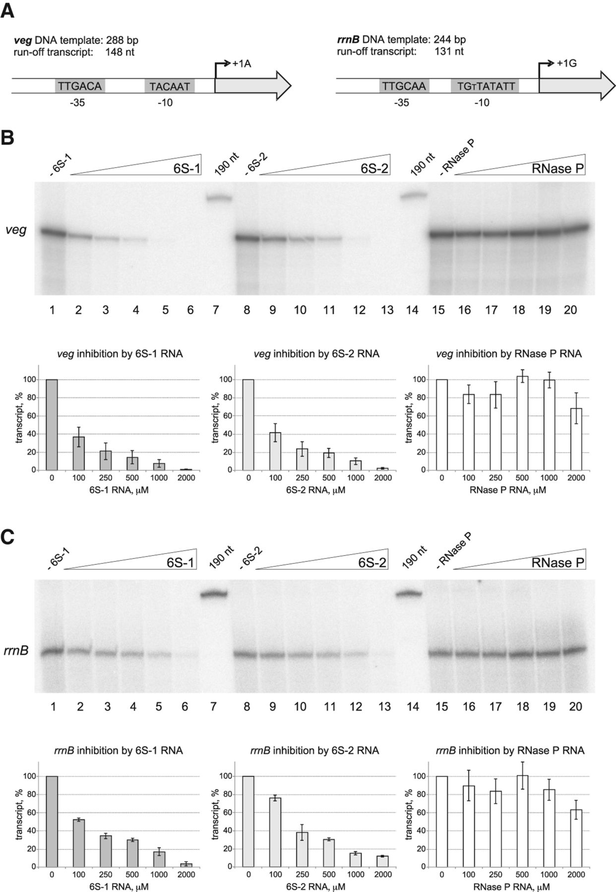
6S-2 RNA as well as 6S-1 RNA inhibit in vitro transcription at the veg and rrnB DNA promoters. (A) Sketch of the two promoter DNA fragments used as transcription templates. The lengths of the T7 run-off transcripts are the theoretical ones based on DNA template complementarity. (B,C) Increasing amounts of 6S-1 RNA (lanes 2–6), 6S-2 RNA (lanes 9–13), or B. subtilis RNase P RNA (RNase P, lanes 16–20, used as a negative control) were added to the (B) veg or (C) rrnB DNA template (f.c. 100 nM) that had been pre-incubated with the σA-RNAP holoenzyme (f.c. 100 nM) in 1× activity buffer. In lanes 1, 8, and 15, no RNA was added. The final RNA concentrations were as follows: 100 nM (lanes 2,9,16), 250 nM (lanes 3,10,17), 500 nM (lanes 4,11,18), 1 µM (lanes 5,12,19), and 2 µM (lanes 6,13,20). Transcription was started by adding the NTP mix containing α-[32P]-UTP. Products were analyzed by 5% denaturing PAGE. 5′-[32P]-end-labeled 6S-1 RNA (190 nt) served as a size marker (lanes 7 and 14). For further details, see Materials and Methods.




