


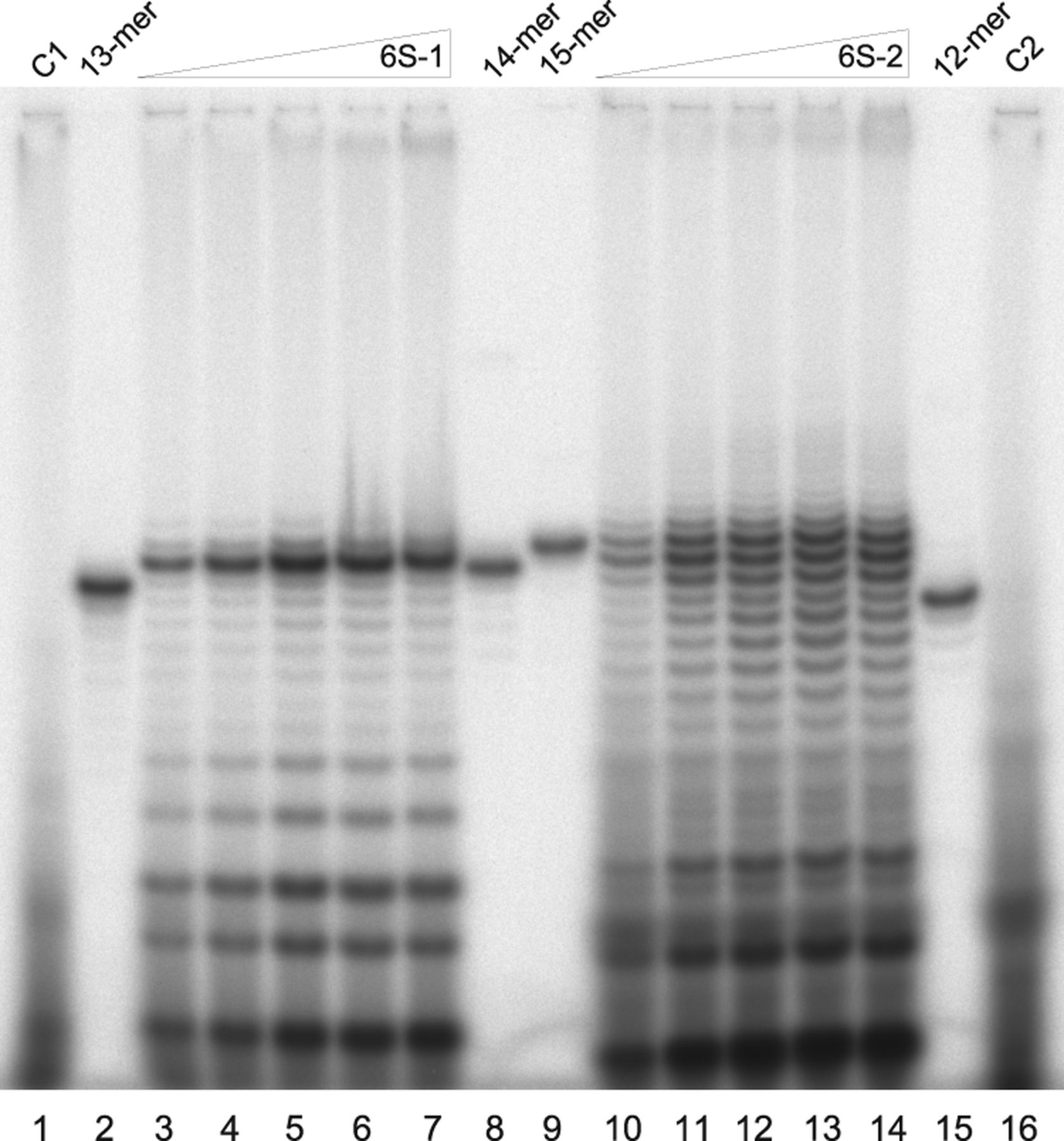
In vitro transcription of pRNAs by the B. subtilis σA-RNAP holoenzyme (1 µM) using increasing amounts of 6S-1 RNA (lanes 3–7) or 6S-2 RNA (lanes 10–14) as templates. The concentration of 6S RNA was 100 nM (lanes 3,10); 500 nM (lanes 4,11); 1 µM (lanes 5,12); 2 µM (lanes 6,13), and 5 µM (lanes 7,14). Products were analyzed by 25% denaturing PAGE. As size markers, 5′-[32P]-end-labeled chemically synthesized pRNA6S-1 13- and 14-mers (lanes 2,8) and pRNA6S-2 12- and 15-mers (lanes 15,9) were used. α-[32P]-UTP was added to transcription in lanes 1 and 3–7, while in lanes 10–14 and 16, α-[32P]-ATP was used for transcript labeling. Lanes 1 and 16 are negative transcription controls where 6S RNA was omitted. Note that synthetic pRNA oligonucleotides used as size standards and carrying a 5′-[32P]-monophosphate may not always comigrate exactly with pRNA transcripts carrying a 5′-triphosphate. For further details, see Materials and Methods.




