


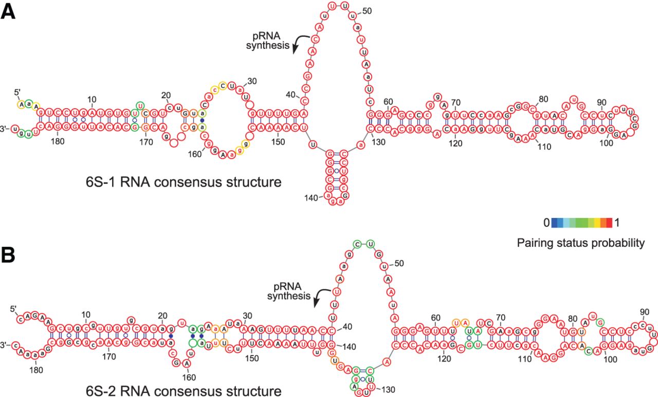
Consensus secondary structures of (A) 14 different 6S-1 and (B) 16 different 6S-2 RNAs according to mlocarna (Will et al. 2007) and RNAalifold (Bernhart et al. 2008) using default parameters. The 6S-1 and 6S-2 RNA species selected for the prediction are derived from Firmicutes, for which two 6S RNAs were identified in their genomes, one falling into the 6S-1 and one into the 6S-2 RNA cluster (for further details, see text and Supplemental Fig. S1). The black arrows depict the experimentally determined starting points for pRNA transcription in B. subtilis (Beckmann et al. 2011). The minimum free energy of the 6S-1 RNA consensus structure is −95.08 kcal/mol and that of 6S-2 RNA, −92.77 kcal/mol. Circle outline colors depict the probability to be unpaired or paired; residues marked in red have a high probability, i.e., their single-stranded or paired state is well-defined. Letters represent the most frequent base at the corresponding alignment position. Red letters in uppercase indicate highly conserved bases with an occurrence of >90%, or >75% indicated by red lowercase letters. Black letters in uppercase indicate nucleotide identities conserved in >50% of the sequences. A small black letter is used for positions with lower or no conservation in the alignment. Empty circles indicate nucleotide insertions in a minor fraction of sequences.




