


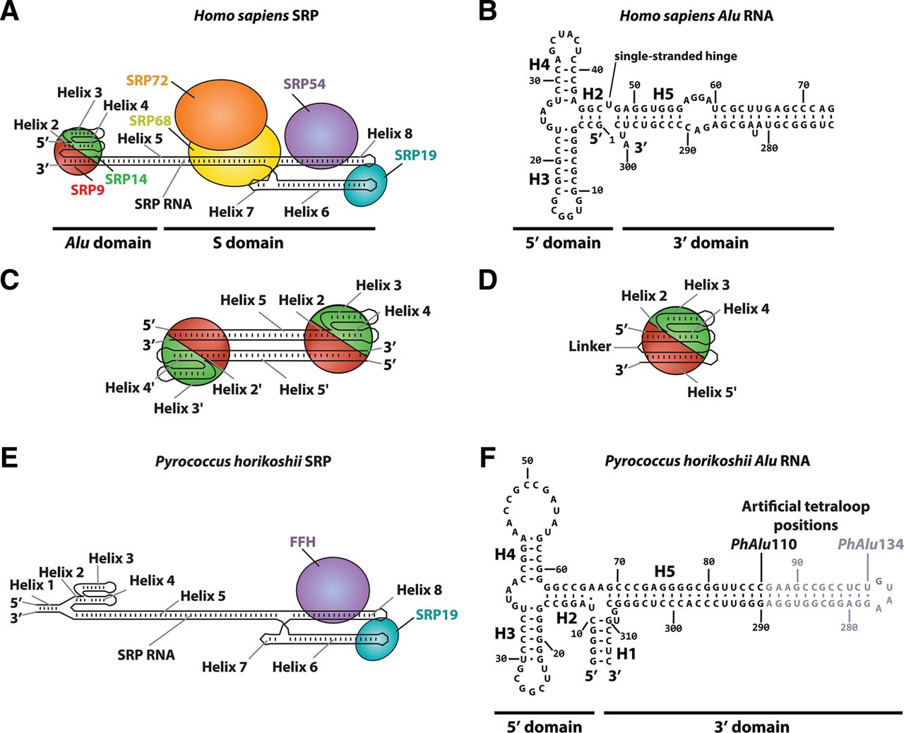
The human and archaeal SRP and Alu domains. (A) Schematic representation of the human SRP showing location of the six proteins (four bound to the S domain and two bound to the Alu domain) and the eight helical segments of the 7SL RNA numbered according to the standard nomenclature of SRP RNAs (Zwieb et al. 2005). SRP proteins are colored as follows: SRP9, red; SRP14, green; SRP68, yellow; SRP72, orange; SRP19, cyan; SRP54/FFH, purple. (B) Sequence of the human Alu RNA with RNA helices marked. Note the single-stranded hinge between the 5′ and 3′ domains. (C) Schematic diagram of the domain-swapped dimeric crystal structure of a rigidly linked human Alu RNA variant (SA86) bound to human SRP9/14 (PDB code 1e8s) (Weichenrieder et al. 2000). Each SRP9/14 heterodimer binds two separate RNA molecules. (D) Hypothetical model of the physiologically active, closed state of the human Alu domain. The single-stranded hinge between the 5′ and 3′ domains allows a single Alu RNA molecule to occupy both binding sites on the SRP9/14 heterodimer (Weichenrieder et al. 2000). (E) Schematic representation of the Pyrococcus horikoshii SRP showing only two proteins bound to the S domain and the additional Helix 1 formed by complementary extensions to the 5′ and 3′ termini. Nomenclature and coloring as in A. The positions of the tetraloops added to close the PhAlu110 and PhAlu134 RNAs are indicated. (F) Sequence of the P. horikoshii Alu RNA with RNA helices marked. Note the three-way junction formed from helices 1, 2, and 5 which rigidifies this region.




