


FIGURE 3.
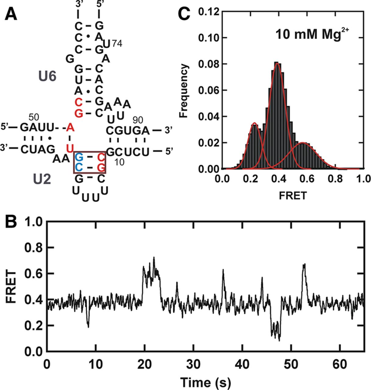
Low FRET state corresponds to the three-helix structure of the U2–U6 complex. (A) Schematic structure of the fourfold mutant hU2–U6 snRNA complex. Residues of U2 and U6 snRNAs participating in helix Ib formation are shown in red. Red box highlights flipped bases (5′ CG-red and 5′ CG-blue) in the U2 stem I. (B) Typical FRET trajectory of the fourfold mutant hU2–U6 snRNA complex. (C) FRET histogram showing the distribution of different FRET states of the mutant U2–U6 snRNA complex in 10 mM Mg2+.




