


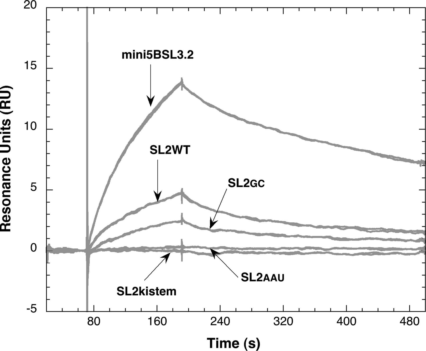
FIGURE 6.
Kinetic analysis of SL2 binding to itself. SL2WT was biotinylated at its 5′ end and immobilized on a streptavidin-coated CM5 sensor chip. SL2 hairpins were injected in duplicate at 500 nM as indicated by the arrows. The experiments were performed at 10°C and at a flow rate of 50 µL/min. The truncated version of 5BSL3.2, mini5BSL3.2, was also injected over the SL2 functionalized surface.




