


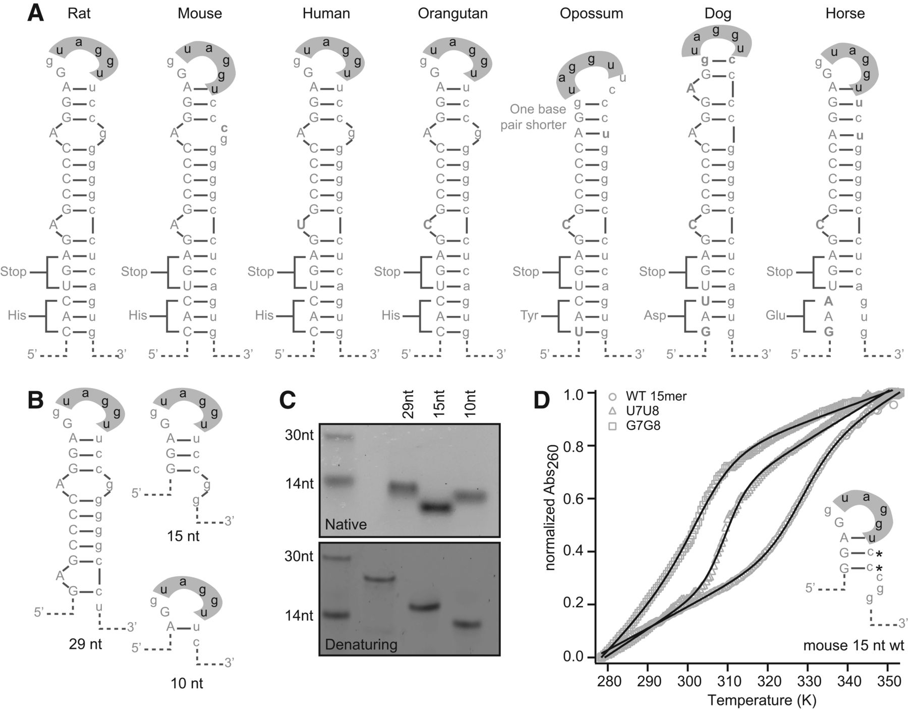
A conserved secondary structure is present at the Mag exon 12 5′ splice site. (A) Putative secondary structure of the region surrounding the splice site from seven vertebrate species. Exon sequence is capitalized, and intron sequence is in lowercase. The hnRNP A1-interacting region is shown in gray. Base pairs are indicated by a horizontal bar. (B) Sequences of the RNAs analyzed in the secondary structure assays in C and D. RNAs are based on the rat sequence. (C) Gel electrophoresis of truncation mutants. RNAs have a higher mobility than expected on a native gel, but migrate according to size on a denaturing gel. (D) Thermal melting analysis of 15 nucleotide RNAs. Mutation of bases predicted to be paired in the stem–loop structure results in a decrease in the melting temperature of the RNA. The predicted structure is presented to the right of the melting curves.




