


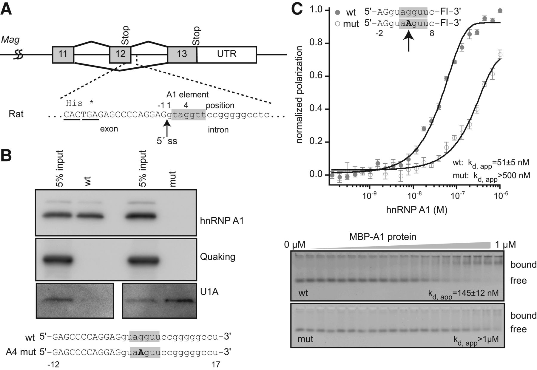
hnRNP A1 interacts with the sequence at the 5′ splice site of Mag exon 12. (A) Diagram of exons 11–13 from rat MAG. Exons are represented with boxes and introns are represented with lines. The sequence surrounding the 5′ splice site is indicated below the diagram. (*) Position of the exon 12 stop codon. Exon sequence is capitalized and intron sequence is lowercase. The putative hnRNP A1-binding element is shaded gray. These conventions are used throughout the remainder of the figures. (B) Streptavidin pulldown assay. Biotin-labeled synthetic RNAs of 29 nucleotides were incubated with a HeLa nuclear lysate, and bound proteins were detected by Western blotting. RNA sequences are presented beneath the image of the blots. (C) Fluorescence polarization and gel electromobility shift analysis of recombinant hnRNP A1 interaction with nine-nucleotide synthetic fluorescent RNAs of the indicated sequence. The apparent dissociation constant for the interaction (Kd, app) is indicated on the figure.




