


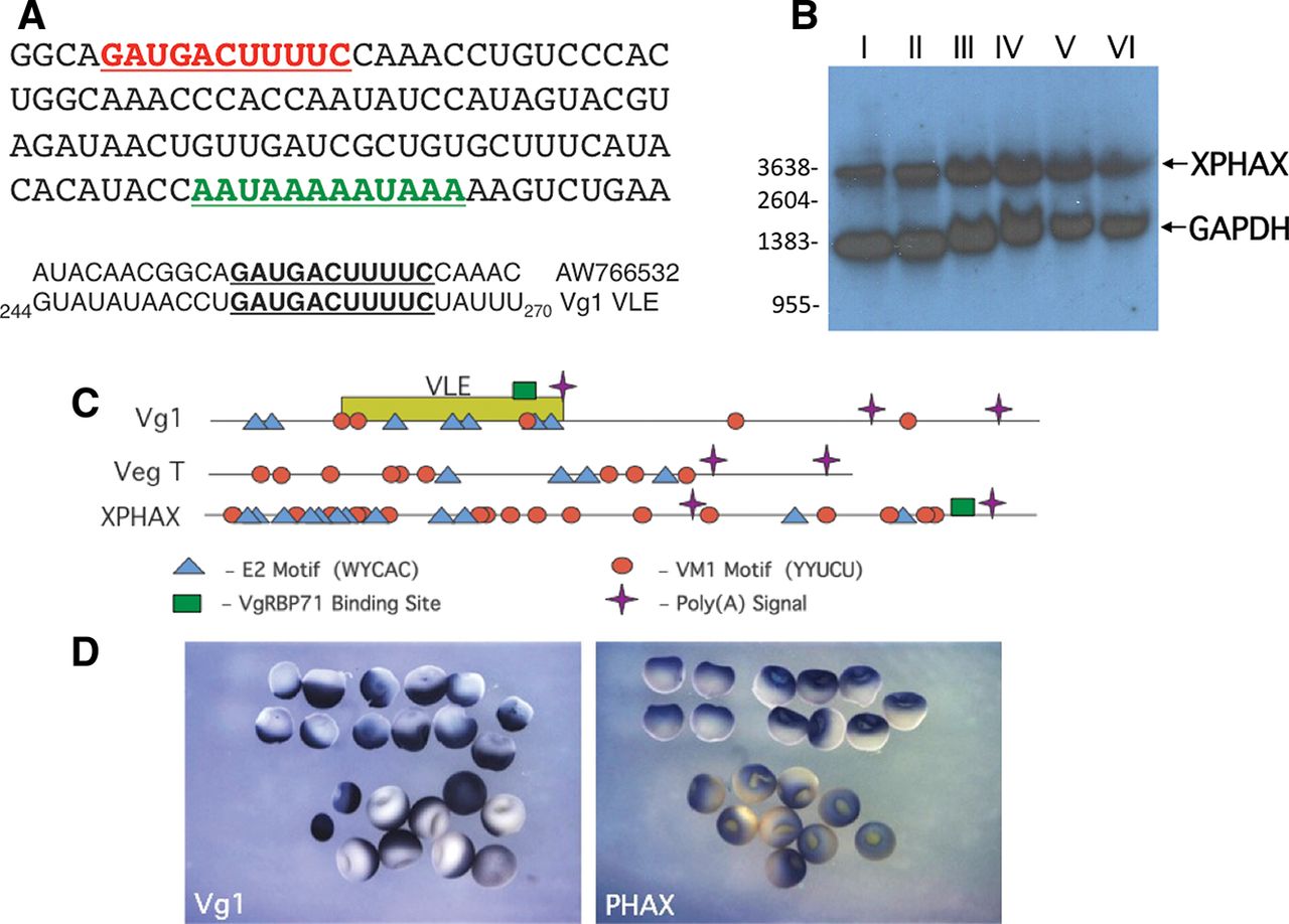
PHAX mRNA is localized to the animal hemisphere of Xenopus oocytes. (A) A portion of the Xenopus EST AW766532 is shown, highlighting the binding site for VgRBP71 (red) and a downstream polyadenylation signal (green). The sequence at the binding site for VgRBP71 (bold, underlined) in the EST is compared with that in Vg1 mRNA. (B) Oocytes were separated according to developmental stage (I–VI) and total RNA isolated for Northern blot analysis. Each lane contains 10 oocyte equivalents of RNA. Complementary probes for PHAX and GAPDH (loading control) mRNAs were used simultaneously. The positions of size standards (nucleotides, nt) are indicated. (C) Scheme showing the relative positions of E2 and VM1 motifs, VgRBP71 binding sites, and polyadenylation signals in the 3′ UTRs of Vg1, VegT, and PHAX mRNAs. (D) Immunohistochemical staining of stage V/VI oocytes following in situ hybridization with digoxigenin-labeled probes for Vg1 mRNA (left panel) and PHAX mRNA (right panel). The discernible indentation on the oocyte surface acts as a marker for the animal hemisphere.




