


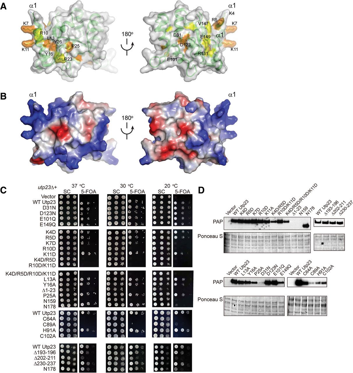
Functional sites of Utp23. (A) Conservation surface of Utp23 PIN domain. Two opposite orientations are shown. Residues that are conserved in >95% and 95%–80% of 134 Utp23 sequences are colored in orange and yellow, respectively. The surface is semitransparent and overlaid on the ribbon representation of Utp23. Some conserved residues are shown as sticks and labeled. (B) Electrostatic potential surface of Utp23 PIN domain shown in the same orientations as in A. The surface is colored from blue to red for positively to negatively charged regions. (C) Yeast growth assay of Utp23 mutants. The utp23Δ haploid strain complemented by a URA3 UTP23 plasmid was transformed with LEU2 plasmids expressing GPD-driven UTP23, no UTP23 (Vector), or the indicated utp23 mutations. Tenfold serial dilutions of the transformants were spotted onto plates containing SC medium or SC with 5-FOA to counter-select for the URA3 UTP23 plasmid and incubated at 37°C, 30°C, or 20°C. (D) Expression of TAP-tagged Utp23 and its mutants from plasmid. Equal amounts of total protein were analyzed by SDS-PAGE, Ponceau S staining, and Western blotting using PAP to detect Utp23-TAP.




