


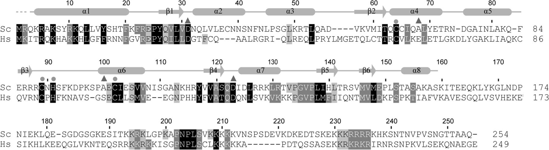
Multiple sequence alignment of Utp23. A total of 134 Utp23 sequences were retrieved from RefSeq with BLAST and aligned with ClustalW and manual adjustment. Only sequences from Saccharomyces cerevisiae (Sc) and Homo sapiens (Hs) are displayed. Residues that are conserved in 95%, 80%, and 70% of all aligned sequences are shaded black, gray, and light gray, respectively. Similarity groups are defined as follows: D and E; K and R; S and T; F, Y, and W; and I, L, M, and V. The secondary structures observed in the crystal structure are indicated on the top. Triangles mark the equivalents to catalytic residues in PIN nucleases and circles denote Zn-coordinating residues.




