


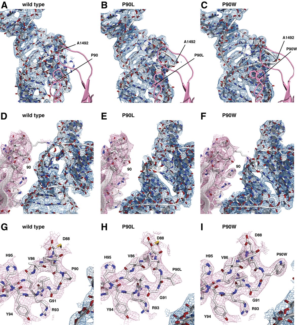
Distortion of the decoding site by SmD mutations in ribosomal protein S12, as observed in composite omit maps contoured at 1σ, of the wild-type (A,D,G), the P90L mutant (B,E,H), and the P90W mutant (C,F,I). (A–C) The decoding site viewed through ribosomal protein S12 (pink). The density for ribosomal protein S12 is excluded from this view for clarity. The position of A1492 is indicated. (D–F) View of the decoding site after an ∼100° rotation about the vertical axis of h44 of the view shown in panels A–C, and showing interaction between S12 and the decoding site. (G,H) Composite omit maps, contoured at 1σ, of ribosomal protein S12 residues 82–96. The densities for the proline and the leucine and tryptophan substitutions at position 90 are clearly visible.




