


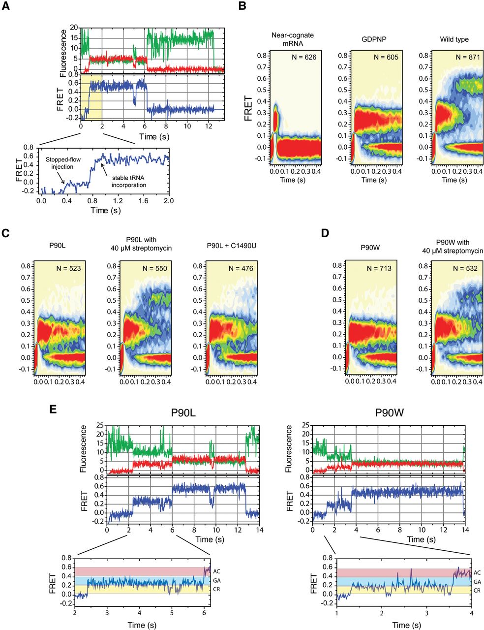
Single-molecule observations of tRNA selection on T. thermophilus 70S ribosomes. (A) Representative single-molecule fluorescence (Cy3 [green]; Cy5 [red]) and FRET trajectories [FRET = ICy5/(ICy3 + ICy5)] obtained during stopped-flow delivery of the ternary complex with Cy5-labeled Phe-tRNAPhe to ribosome complexes containing Cy3-labeled P-site fMet-tRNAfMet. The first 2 sec of the FRET trajectory (yellow) is expanded beneath, showing clear progression from zero-FRET to a fully accommodated (AC), high-FRET state via on-path intermediates in the tRNA selection process. (B) Post-synchronized FRET trajectories combined in two-dimensional contour plots from wild-type ribosomes decoding near-cognate UCU codon (left), cognate UUC codon with GTP replaced by the nonhydrolyzable GTP analog GDPNP (middle), and cognate UUC codon (right). (C) Decoding by P90L ribosomes (left), P90L ribosomes in the presence of 40 μM streptomycin (middle), and P90L/C1490U SmI mutant ribosomes (right). (D) tRNA incorporation by P90W ribosomes in the absence (left) or presence (right) of 40 μM streptomycin. (E) Representative traces state from P90L (left) and P90W (right) complexes showing molecules that successfully reached the fully accommodated (AC) state after fluctuating reversibly between FRET states assigned to codon recognition (CR) and GTPase-activated (GA) states. Fluorescence and FRET trajectories are shown as described in Figure 1A.




