


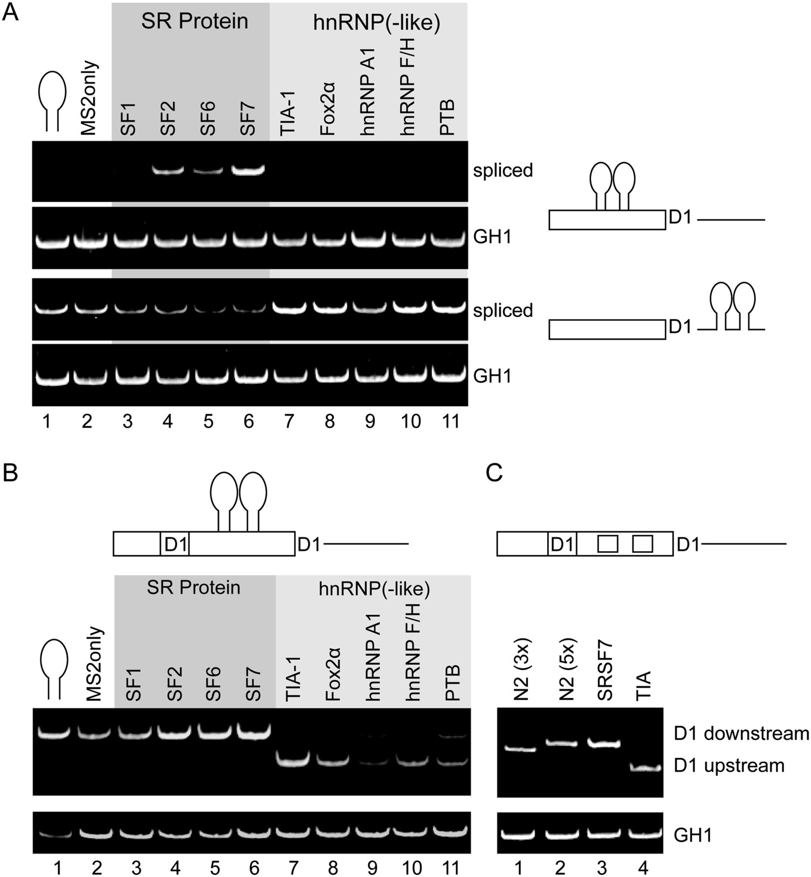
MS2 tethering of splicing regulatory proteins mimics positional requirements for 5′ss activation. (A) Expression of MS2 fusion proteins results in position-dependent activation or repression of splicing using reporters that harbor MS2 binding sites upstream of or downstream from the D1 5′ss. Comparable expression of all MS2 fusion proteins was verified by Western blot analysis (Supplemental Fig. 5). (B) Schematic diagram of the splicing reporter in which the two MS2 binding sites are flanked by the identical up- and downstream 5′ss D1. The splicing pattern of each sample was analyzed using semi-quantitative RT-PCR analysis. (C) Identical experiments were performed using splicing reporters where the MS2 binding sites were replaced by high-affinity binding sites for endogenous TIA-1 and SRSF7.




