


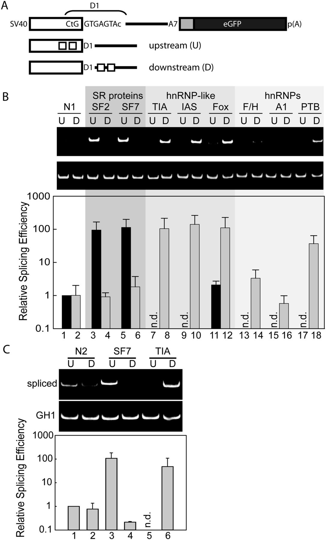
The positioning of splicing regulatory elements determines their effect on 5′ splice site (5′ss) activation. (A) Schematic drawing of the RNA splicing reporter. Specific binding sites for splicing regulatory proteins are immediately upstream of or downstream from the D1 5′ss, as indicated by boxes. HeLa cells were transiently transfected with each of the reporter constructs and analyzed by semi-quantitative and real-time PCR to determine the position-dependent activity of SREs (B) and to demonstrate that each SRE displays position-dependent activation and repression of splicing (C). N1 and N2 are splicing neutral and near neutral control sequences (Supplemental Fig. 2). U and D refer to upstream and downstream positions relative to the D1 5′ss. n.d. refers to not detected above background. All data were normalized to the constitutively spliced GH1 mRNA.




