


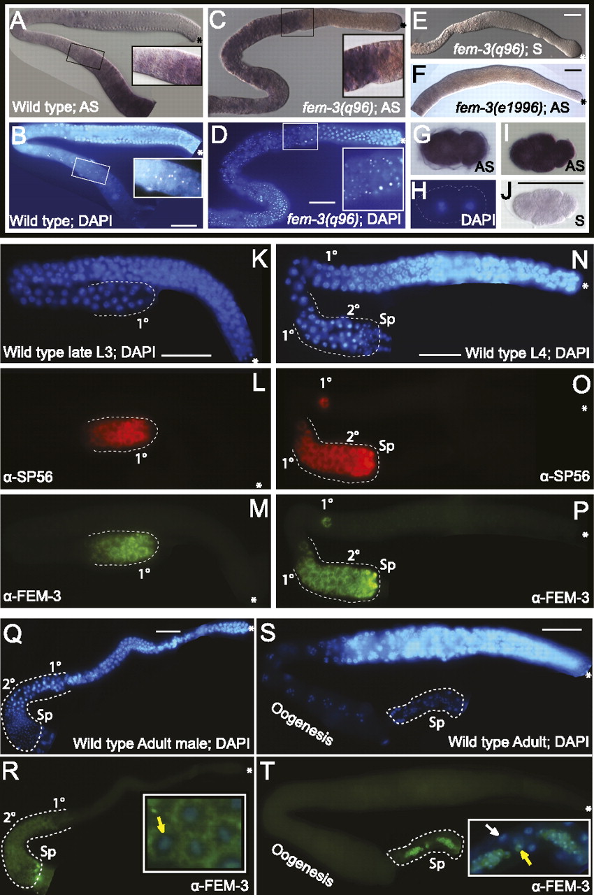
Expression of fem-3 in embryos and germlines. (A) In situ localization of fem-3 mRNA in adult germlines. (B) Nuclear DAPI staining; region with oocytes in diplotene is boxed and magnified. (C,D) In situ hybridization of fem-3 in an adult fem-3 gain-of-function mutant germline. Boxed: primary and secondary spermatocytes. The proximal portion (to the left of the box) of the gonadal arm is filled with spermatids. (E) Germline hybridized with a fem-3 sense RNA probe. (F) No signal was observed in a fem-3(e1996, null) mutant germline. (G,H) Maternal fem-3 mRNA in a two-blastomere embryo and staining with DAPI. (I) fem-3 mRNA was found in all blastomeres of a four-cell embryo: ABa (left), ABp (top), EMS (bottom), and P2 (right). (J) Staining of a four-cell embryo with a fem-3 sense RNA probe. FEM-3 protein localizes to the sperm lineage: (K–M) Wild-type L3 germline stained with DAPI, SP56 anti-sperm and anti-FEM-3 antibodies. The region outlined contains primary (1°) spermatocytes. (N–P) Staining of a wild-type germline of larval stage L4: (2°) secondary spermatocytes, (Sp) spermatids. (Q,R) Distribution of FEM-3 in an adult male germline. The region containing spermatocytes and spermatids is outlined. The inset shows high magnification of spermatids stained for DNA and FEM-3. The expression of FEM-3 is clearly cytoplasmic. (S,T) Localization of FEM-3 in a dissected adult hermaphrodite germline: FEM-3 is only present in the cytoplasm of spermatids. Inset: magnification of spermatids, merged DAPI, and anti-FEM-3 stainings. Yellow arrow, nucleus of spermatid. White arrow, nucleus of somatic cell of the spermatheca. Asterisks indicate the distal end of the germline. Bar, 10 μm.




