


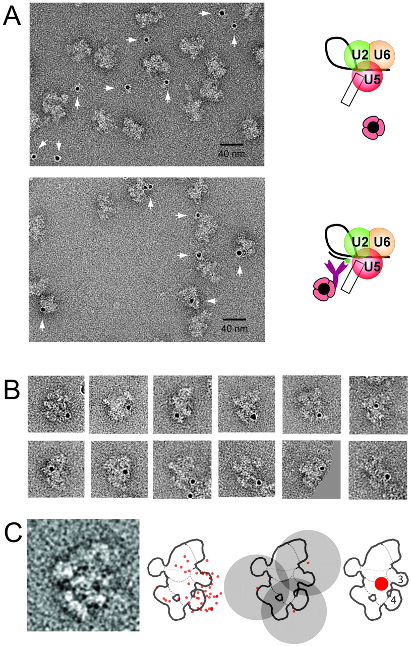
EM localization of an intron position close to the anchoring site in the C complex. Labeling was as described in Fig. 3, except that here the biotinylated “label intron anchoring site” oligonucleotide was used. (A) General field of C complexes incubated with PAG either in the absence (upper panel) or presence (lower panel) of a biotinylated oligonucleotide complementary to the intron just upstream of the anchoring site. (B) Gallery of PAG-labeled C complexes seen in the dominant view. (C) Left: Superposition of all 49 gold-labeled C complexes used for locating the intron position close to the anchoring site, showing the dominant projection. As in Figs. 3C and 4C, all of the typical features are seen. Second from left: Outline sketch of the dominant view with all gold positions indicated. Third from left: An example of the use of the 17-nm rule for three gold labels. Far right: Using the 17-nm rule for all 49 gold labels, the biotin-labeled nucleotide positioned on the intron close to the anchoring site localizes within the red area close to domains 3 and 4 (labeled) of the C complex.




