


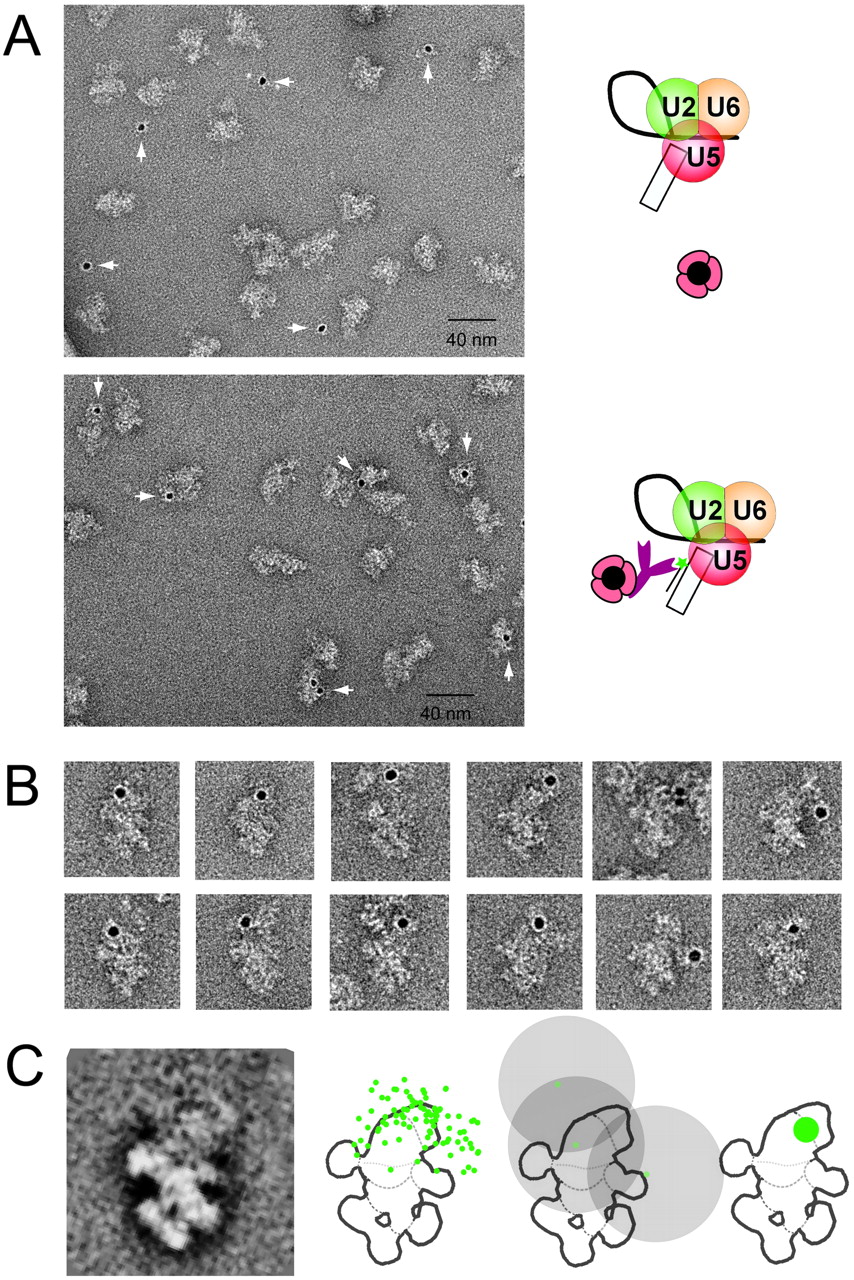
EM localization of an exon 1 position close to the cleaved 5′ splice site in the C complex. Assembled human C complexes were treated successively with biotinylated RNA (“label oligo exon 1”) (Fig. 2B) and antibiotin IgG, then stabilized by the GraFix method, incubated with protein A-coated colloidal gold (PAG), and examined by EM using negatively stained samples (see Materials and Methods). (A) General field of C complexes incubated with PAG either in the absence (upper panel) or presence (lower panel) of the biotinylated oligonucleotide complementary to exon 1. (B) Gallery of PAG-labeled C complexes seen in the dominant view. (C) Superposition of all 87 gold-labeled C complexes used for localization of exon 1, showing the dominant projection (left). All typical structural features of the dominant view can be recognized. The “head” (domain 2) appears blurred as many gold particles are found there (see text). In the schematic outline drawing of the C complex (second from left), all of the positions of gold particles in the C complex images showing the dominant view are indicated. The sketch second from the right shows, as an example, 17-nm circles around three gold particles and the overlap region to which the epitope is assigned. Using the 17-nm rule for all 87 gold labels (Wolf et al. 2009), the biotin of the oligonucleotide positioned approximately in the middle of exon 1 (48 nt upstream of the 3′ end) can be localized to the green area in the head of the C complex (right).




