


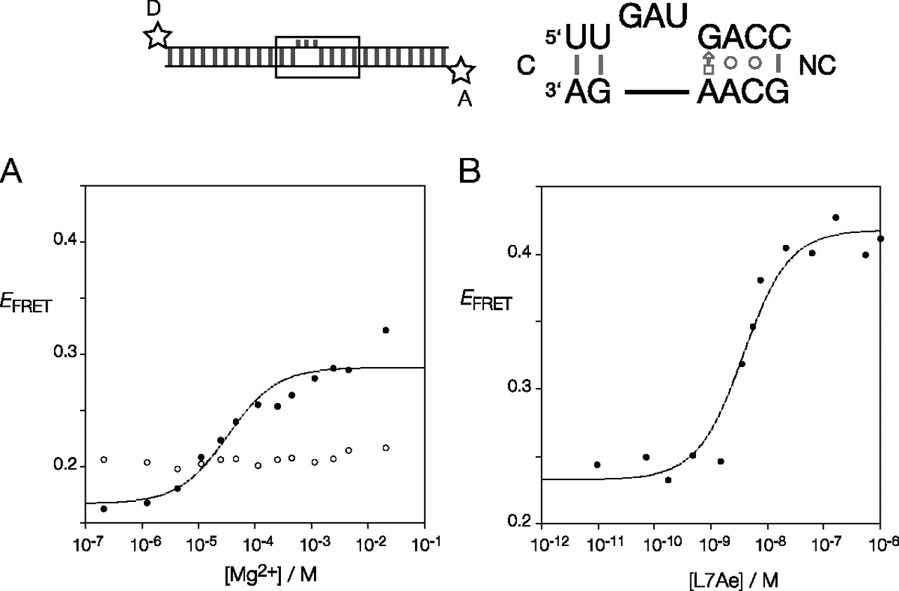
FIGURE 2.
FRET analysis of the folding of T. solenopsae Kt-23 induced by addition of Mg2+ ions and by L7Ae binding. The k-turn sequence was centrally located within a 25-bp RNA duplex terminally labeled with fluorescein (donor, D) and Cy3 (acceptor, A) fluorophores. FRET efficiency was measured in the steady-state. (A) Mg2+ ion-induced folding of the natural T. solenopsae Kt-23 (closed circles) and the L1 2′H variant (open circles). The change in EFRET was fitted using Equation (1). (B) L7Ae-induced folding of the natural T. solenopsae Kt-23. The change in EFRET was fitted using Equation (2).




