


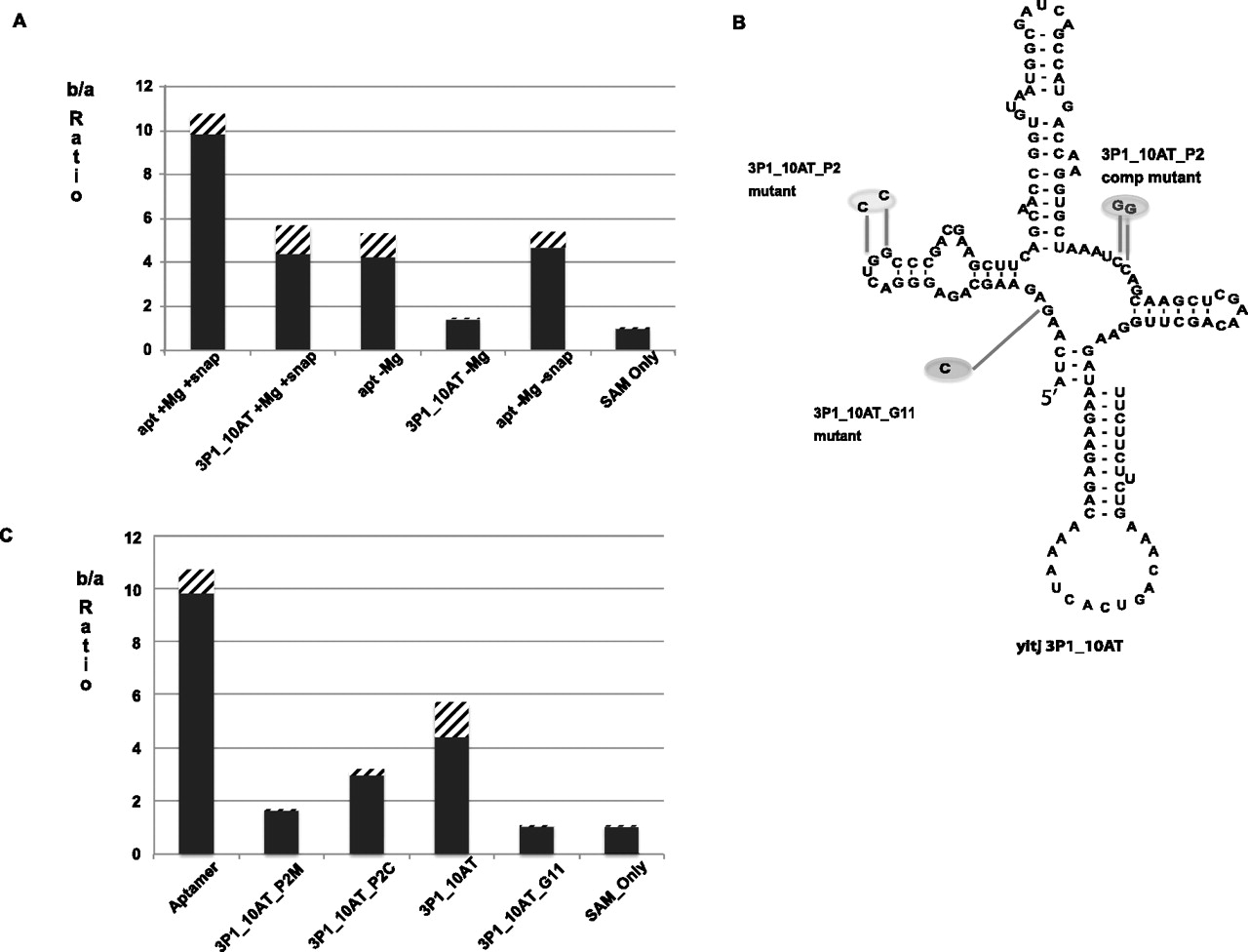
(A) Ratio of number of counts in chamber b to number of counts in chamber a in equilibrium dialysis for B. subtilis yitJ SAM-I riboswitch aptamer and 3P1_10AT hybrid in the presence (+Mg) and absence (−Mg) of Mg2+ and with (+snap) and without (−snap) snap cooling. (B) Schematic representation of B. subtilis yitJ SAM-I riboswitch 3P1_10AT pseudoknot and G11 mutants. (C) Equilibrium dialysis data for Aptamer, Pseudoknot mutant (3P1_10AT_P2M), pseudoknot compensatory mutant (3P1_10AT_P2C), 3P1_10AT, and 3P1_10AT_G11 mutant. Equilibration of SAM in the two chambers is shown with SAM only. The error bars (stripes) represent the standard deviation for at least three replicates for each experiment.




