


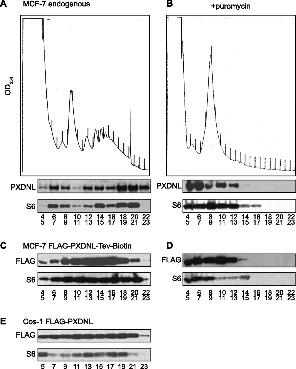
Polysome profile analysis of 57-kDa PXDNL. (A) Cytoplasmic extract from MCF-7 cells was separated on a 10%–50% sucrose gradient. The upper panel shows the absorbance at 254 nm during fractionation. Every two fractions starting with fraction 4 were pooled and ethanol precipitated, and endogenous protein was identified by Western blotting with anti-PXDNL antibody or antibody to ribosomal protein S6. (B) MCF-7 cells were treated with puromycin for 15 min prior to lysis and analyzed as in A. (C) Tetracycline repressor was introduced by viral transduction into MCF-7 cells. These were stably transfected with a tetracycline-inducible plasmid expressing the same form of PXDNL as in Figure 3B with an N-terminal Flag tag and a C-terminal sequence that is biotinylated in vivo. Cells were induced for 6 h with doxycycline, and cytoplasmic extract was analyzed on a linear 10%–50% sucrose gradient as in A with the exception that Western blotting of the recovered gradient fractions was performed using anti-Flag monoclonal antibody and with antibody to ribosomal protein S6. (D) The same cells that were used in C were treated for 15 min with puromycin prior to lysis and analyzed as in C. (E) Cos-1 cells were transiently transfected with a plasmid expressing 57-kDa PXDNL with just an N-terminal Flag tag. Cytoplasmic extract was separated on a linear 10%–50% sucrose gradient as in A and C, and protein recovered in odd-numbered fractions was detected by Western blotting with anti-Flag monoclonal antibody.




