


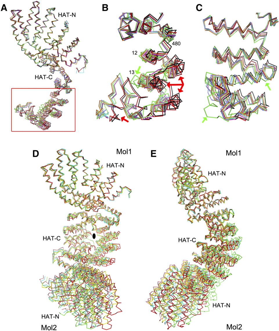
Conformational variability of the K. lactis HAT domain dimer. (A) Overlay of the nine HAT domain monomers in the three different crystal forms. Residues 28–480 are superposed for this overlay. The red box highlights the structural differences in the C-terminal segment (residues 481–582) of the monomers. (B) Close-up view of the structural differences in the C-terminal region. Red arrows point to large differences for two of the monomers (red and black) of a HAT domain dimer in crystal form 3. Green arrows point to large differences for the monomer in crystal form 1 (green). (C) Panel B viewed after a 90° rotation around the vertical axis. (D) Overlay of the five HAT domain dimers in the three different crystal forms. Residues 28–480 in the first monomer are superposed for this overlay. Large differences in the positions of the second monomer, especially the HAT-N domain, are clearly visible. (E) Panel D viewed after a 90° rotation around the vertical axis.




