


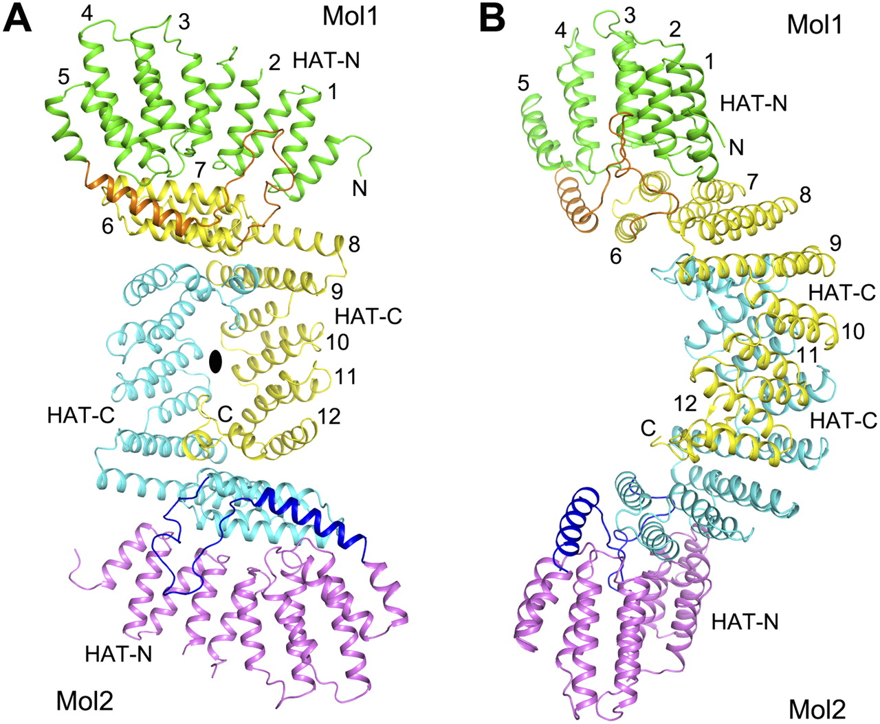
FIGURE 2.
Structure of the HAT domain dimer of K. lactis Rna14. (A) Schematic drawing of the structure of the HAT domain dimer, viewed down the twofold axis (black oval). The HAT-C domain of one monomer is shown in yellow; the other, in cyan. The HAT-N domain of one monomer is shown in green; the other, in magenta. The linker between the HAT-N and HAT-C domains is shown in orange and blue, respectively. (B) Structure of the HAT domain dimer, after 90° rotation around the vertical axis from panel A. All structure figures were produced with PyMOL (www.pymol.org).




