


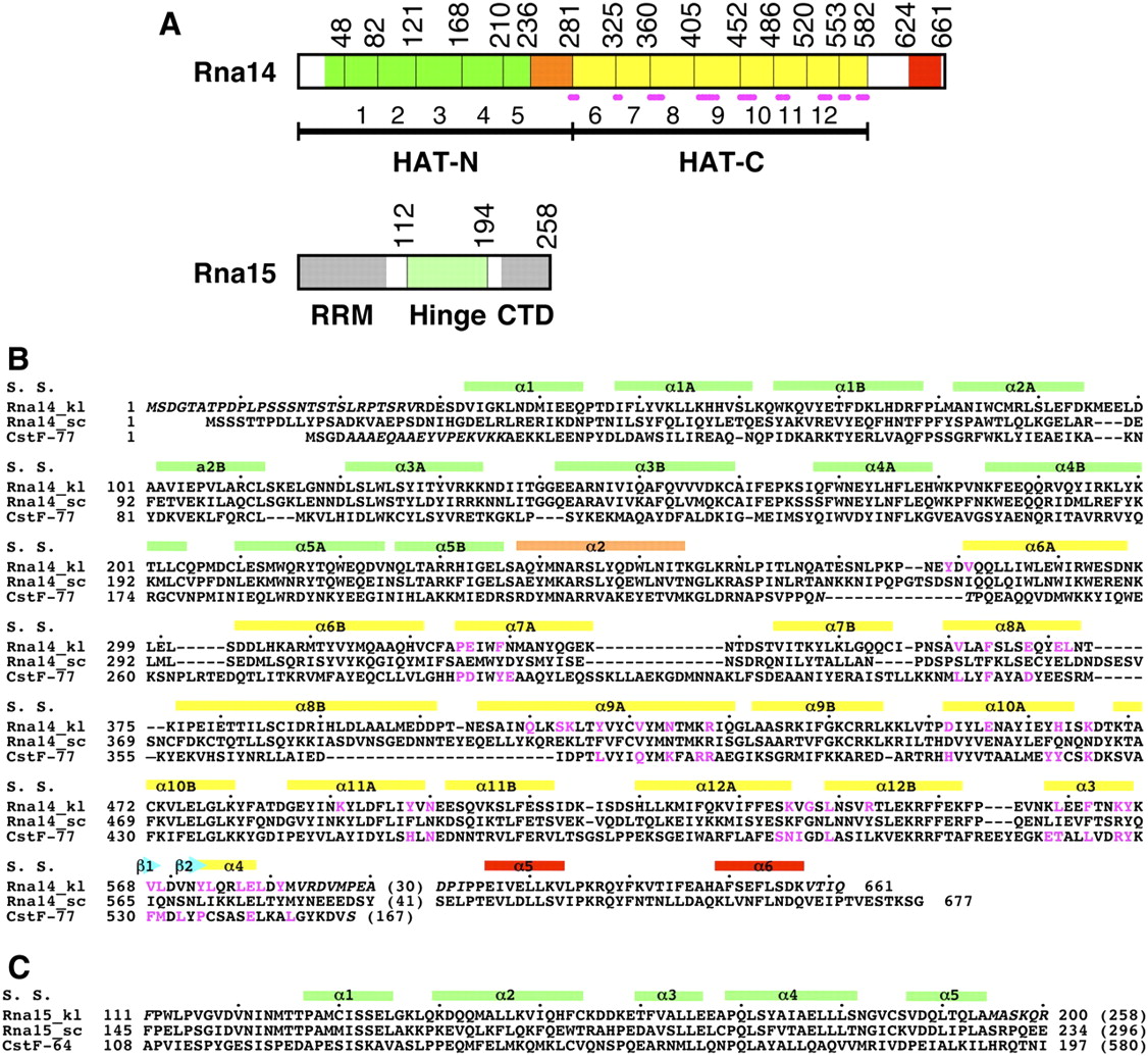
Sequence alignments of Rna14 and Rna15. (A) Schematic drawing of the domain organization of K. lactis Rna14 and Rna15. Motifs in the HAT-N domain of Rna14 are shown in green and labeled; those in the HAT-C domain, in yellow. The linker between the two domains is shown in orange. The dots in magenta indicate residues in the dimer interface of the HAT domain. The C-terminal region that interacts with Rna15 is shown in red. The hinge region of Rna15 is shown in light green; the RNA recognition module (RRM) and the C-terminal domain (CTD), in gray. (B) Structure-based sequence alignment of K. lactis Rna14 (Rna14_kl), yeast Rna14 (Rna14_sc), and murine CstF-77. The secondary structure elements (S.S.) are labeled. Residues shown in magenta are in the dimer interface of the HAT domain. Residues missing in the crystal structures are shown in italics. (C) Sequence alignment of the hinge region of K. lactis Rna15 (Rna15_kl), yeast Rna15 (Rna15_sc), and murine CstF-64. The total number of residues for each protein is indicated in the parenthesis.




