


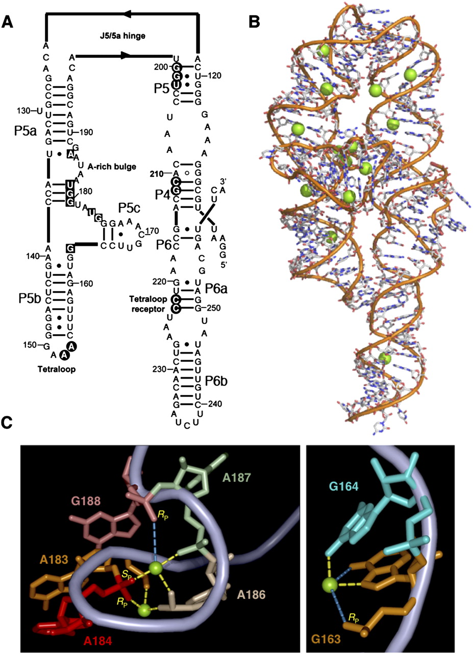
Structure of ΔC209 P4–P6 RNA. (A) Secondary structure of ΔC209 P4–P6. Nucleotides protected from hydroxyl radical cleavage in the folded state are shown as black circles (tertiary protections) or as black squares (subdomain protections). (B) Crystal structure of ΔC209 P4–P6 (derived from PDB file 1HR2). Green spheres represent magnesium ions observed in the structure. (C) Detail of metal ion binding sites in ΔC209 P4–P6 relevant to this work. Within the A-rich bulge (left) and the trihelical junction (right), magnesium ions make several inner-sphere (yellow dashes) and outer-sphere (blue dashes) contacts to nucleobases and nonbridging phosphate oxygens. The specific phosphate oxygens modified in this work are labeled in yellow text.




