


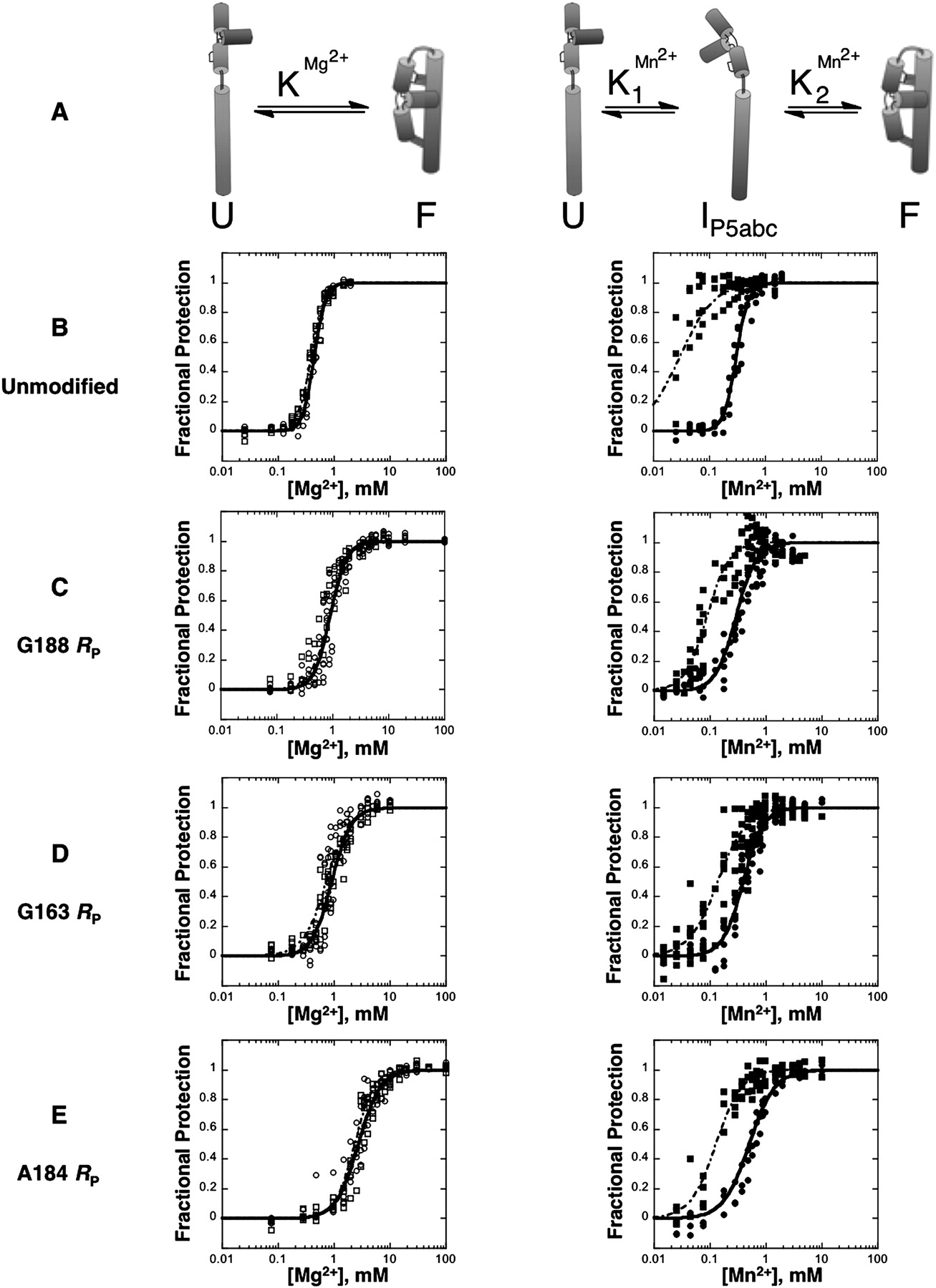
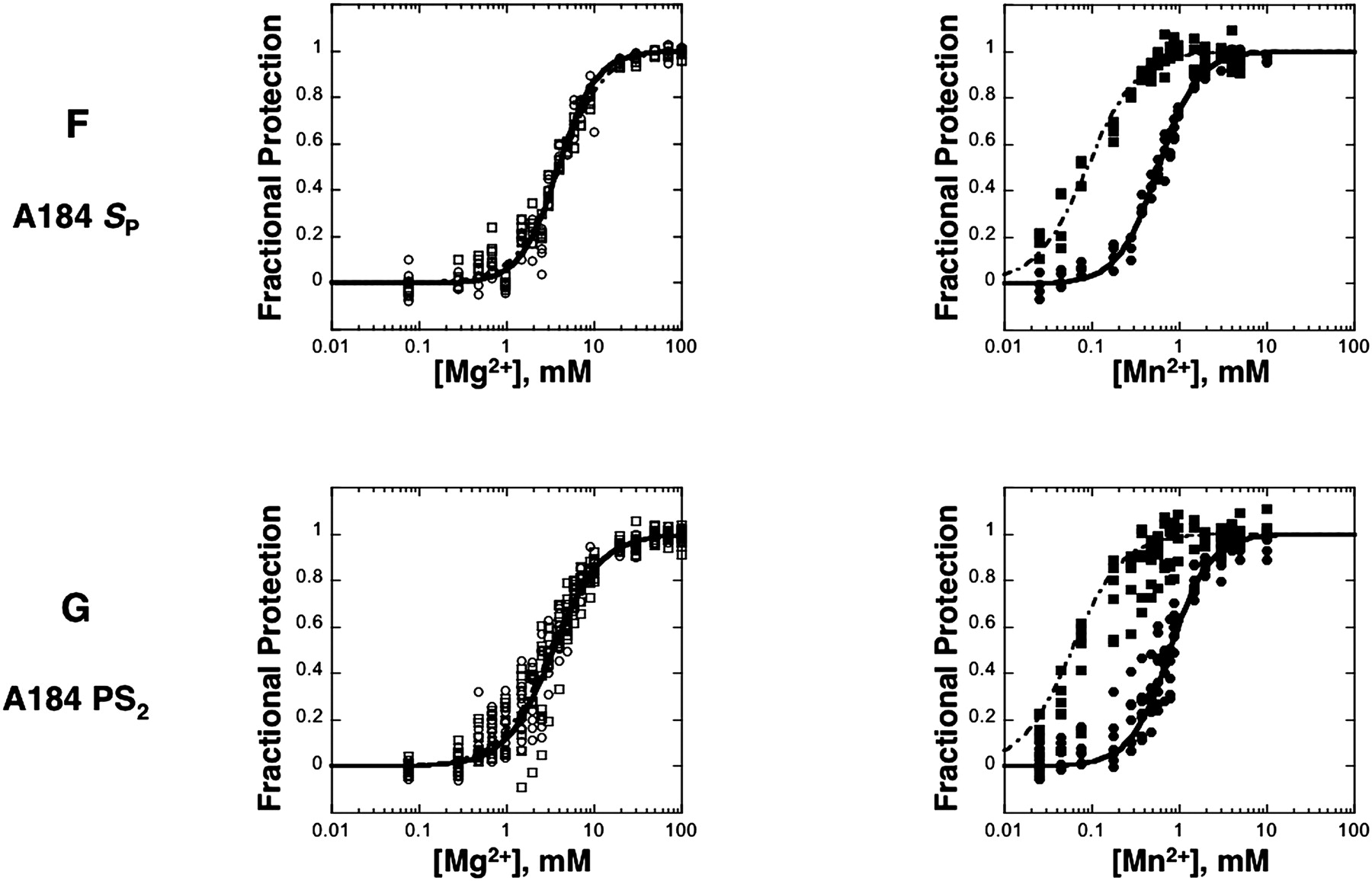
Mg2+- or Mn2+-induced folding of P4–P6 variants in a background of 20 mM Na+. Folding of unmodified P4–P6 in Mg2+ follows an apparent two-state transition modeled as U-to-F (A, left), in which both subdomain protections (B–G, left, open squares and dashed curves) and tertiary protections (B–G, left, open circles and solid curves) appear over approximately the same Mg2+ concentration range. In contrast, folding in Mn2+ follows the three-state transition U-to-IP5abc-to-F (A, right), in which the subdomain protections (B–G, right, closed squares and dashed curves) appear at lower concentrations than the protections associated with formation of tertiary structure (B–G, right, closed circles and solid curves). Data points and curve fits were obtained for the subdomain and tertiary protections analogously to the procedure described for Figure 3.




