


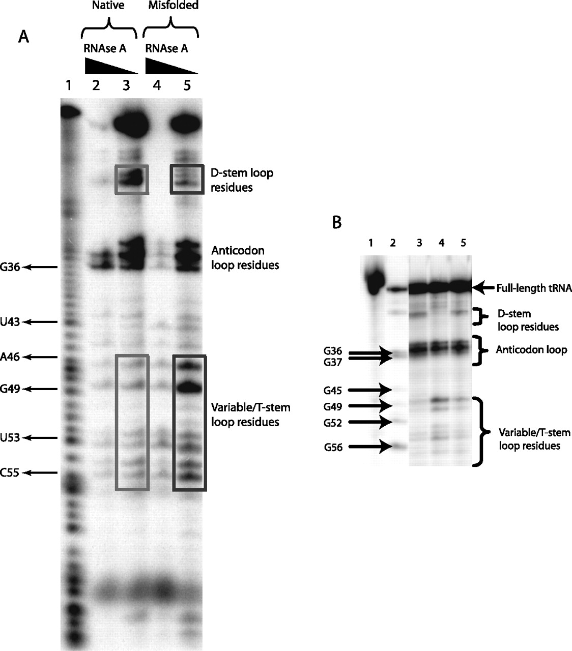
(A) RNase A footprinting of native and non-native forms of MT tRNAGln(CUG). Lane 1 corresponds to an alkaline hydrolysis ladder. MT tRNAGln(CUG) was either heat-denatured and slow cooled following Mg2+ addition to generate native species (lanes 2,3) or heat-denatured and flash-cooled to 0°C with Mg2+ addition to generate non-native species (lanes 4,5). Lanes 2 and 4 represent native and non-native forms of tRNAs, respectively, that were subjected to RNase A digestion under conditions (1 ng/mL enzyme) allowing for complete digestion. In contrast, lanes 3 and 5 represent native and non-native forms of tRNAs, respectively, that were subjected to RNase A digestion under conditions (0.1 ng/mL enzyme) allowing for partial digestion. Differences in the intensities of RNase A digestion patterns, observed in the T- and D-arms, are boxed. (B) RNase S1 footprinting of native and non-native forms of MT tRNAGln(CUG). (Lane 1) Full-length tRNA; (lane 2) T1 nuclease ladder; (lane 3) native tRNA formed by heating to 80°C followed by slow-cooling in the presence of MgCl2; (lane 4) non-native tRNA generated by heating to 80°C and flash-cooling to 0°C in the presence of MgCl2; and (lane 5) native tRNA generated by heating to 80°C, flash cooling in the presence of MgCl2 at 0°C for 5 min, and subsequent refolding at 21°C for 2 h. The concentration of S1 nuclease used in the experiments shown in lanes 3 through 5 is 3 units per milliliter, calibrated to provide partial digestion of the substrate (data not shown).




