


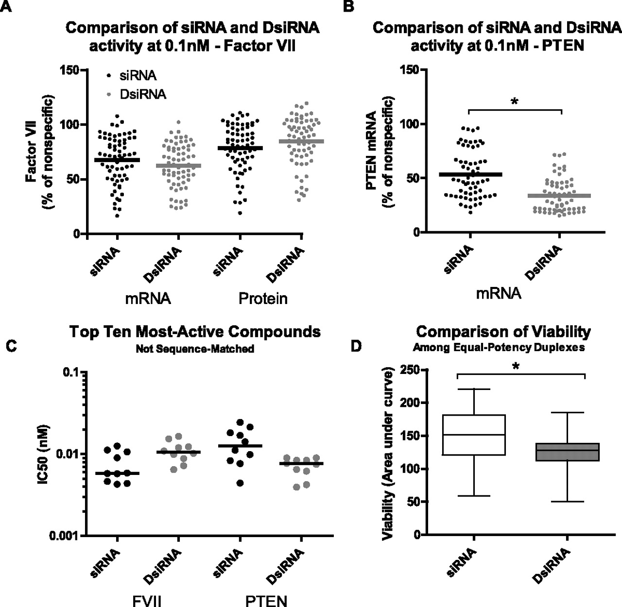
In vitro activity and viability. (A,B) HeLa cells stably transfected with FVII (A) or parental HeLa cells (B) were reverse-transfected using Lipofectamine RNAiMax with 0.1 nM duplex. FVII protein (A) was quantified by chromogenic assay, while mRNA (A–C) was quantified by branched DNA assay. Values were normalized to percent of nonspecific 21-mer control. (C) Transfection and assay as in A and B, but a sixfold dilution series containing eight points was prepared, ranging from 20 nM to 75 fM. After normalization to nonspecific 21-mer, IC50 values were determined. Log-transformed IC50 values were compared by a two-tailed Student's t-test. (D) Viability assay in HeLa using sequence-matched compounds of equal efficacy targeting FVII (six pairs) or PTEN (six pairs). Reverse transfection was performed with Lipofectamine RNAiMax and 5 nM duplex. Samples were evaluated by CellTiter Blue on Days 2, 3, and 4 after transfection. Data were normalized to mock-transfected, and area under the curve (AUC) values were calculated as a measure of cumulative viability. Data were compared by two-way ANOVA (effects of structure and sequence). Data (A–D) are averages of at least two independent experiments, each with a minimum of two biological replicates. (*) p < 0.05.




