


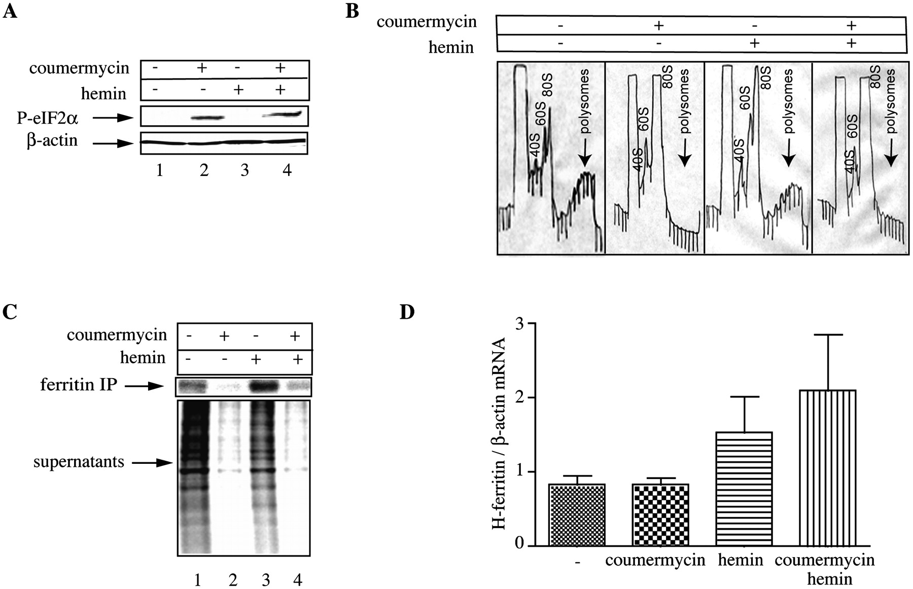
De novo ferritin synthesis despite global translational arrest upon eIF2α phosphorylation. HT1080-GyrB-PKR cells were treated with coumermycin (100 ng/mL) and hemin (100 μM) for 6 h. (A) Cell lysates were analyzed by Western blotting with an eIF2α phosphospecific (top) and a β-actin antibody (bottom). (B) Cytoplasmic extracts were fractionated on sucrose gradients with continuous monitoring of A260; arrows indicate the heavy polysomes. (C) The cells were metabolically labeled with [35S]methionine/cysteine for 2 h and 1000 μg of cell extracts were subjected to immunoprecipitation with 30 μg of ferritin antibody. Immunoprecipitated material and the IP supernatants were analyzed by SDS-PAGE on a 12% gel. The ferritin bands and the lanes showing global protein synthesis were visualized by autoradiography. (D) Analysis of H-ferritin mRNA from whole-cell lysates by qPCR.




