


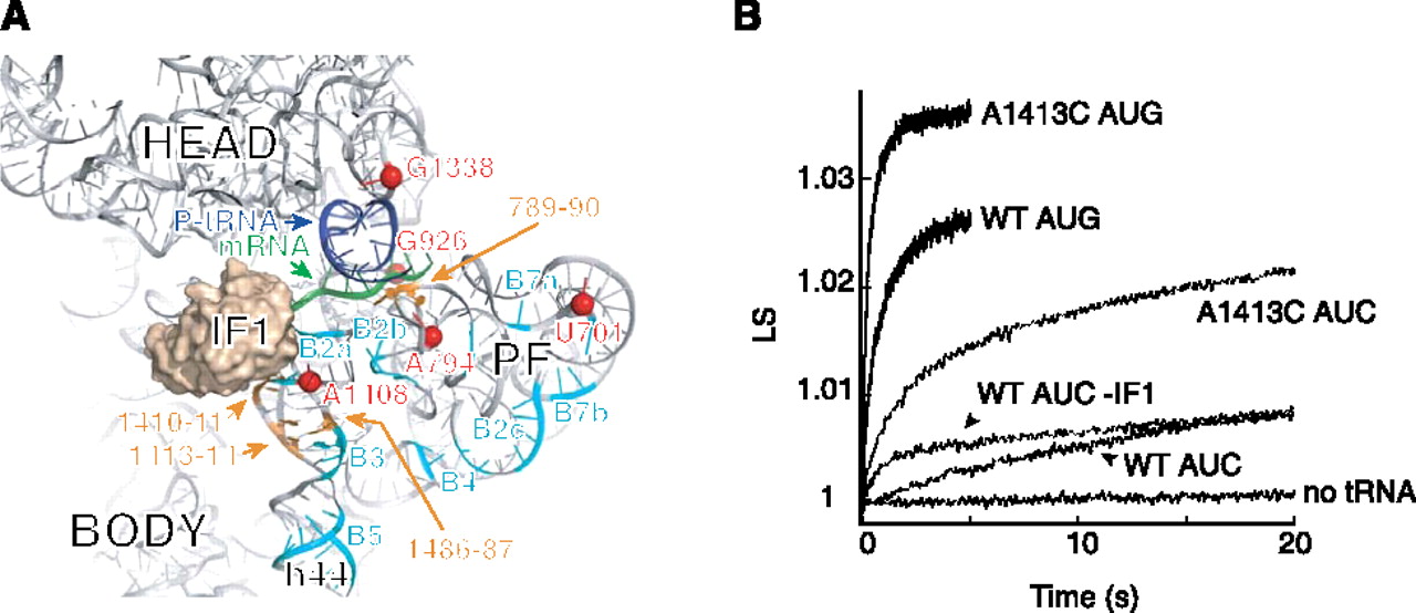
Mutation A1413C stimulates 70SIC formation. (A) Tertiary structure of 16S rRNA of the 30S subunit (ribosomal proteins are omitted for clarity), viewed from the subunit interface. The positions of IF1 (tan), P-site tRNA (dark blue), mRNA (green), and nucleotides contributing to intersubunit bridges (cyan, as indicated) are shown. (Orange) Positions of mutations that increase translation from noncanonical start codons (Qin and Fredrick 2009). (Red spheres) Nucleotides identified in this study that exhibit different chemical reactivity in 30SIC(AUG) versus 30SIC(AUC). This image was generated using PDB files 2J02 and 1HR0 (Carter et al. 2001; Selmer et al. 2006). (B) Examples of experiments measuring apparent rates of 50S docking. Control (WT) or mutant (A1413C) 30S subunits (0.075 μM) were pre-incubated for 30 min at 37°C with initiation factors (0.2 μM each), GTP (100 μM), fMet-tRNA (0.2 μM), and mRNA (with start codon AUG or AUC as indicated, 0.5 μM). Then, each 30S complex was rapidly mixed with an equal volume of 50S subunits (0.23 μM) in a stopped-flow apparatus, and scattered light was measured as a function of time. Ordinate values correspond to relative units of light scattering (LS).




