


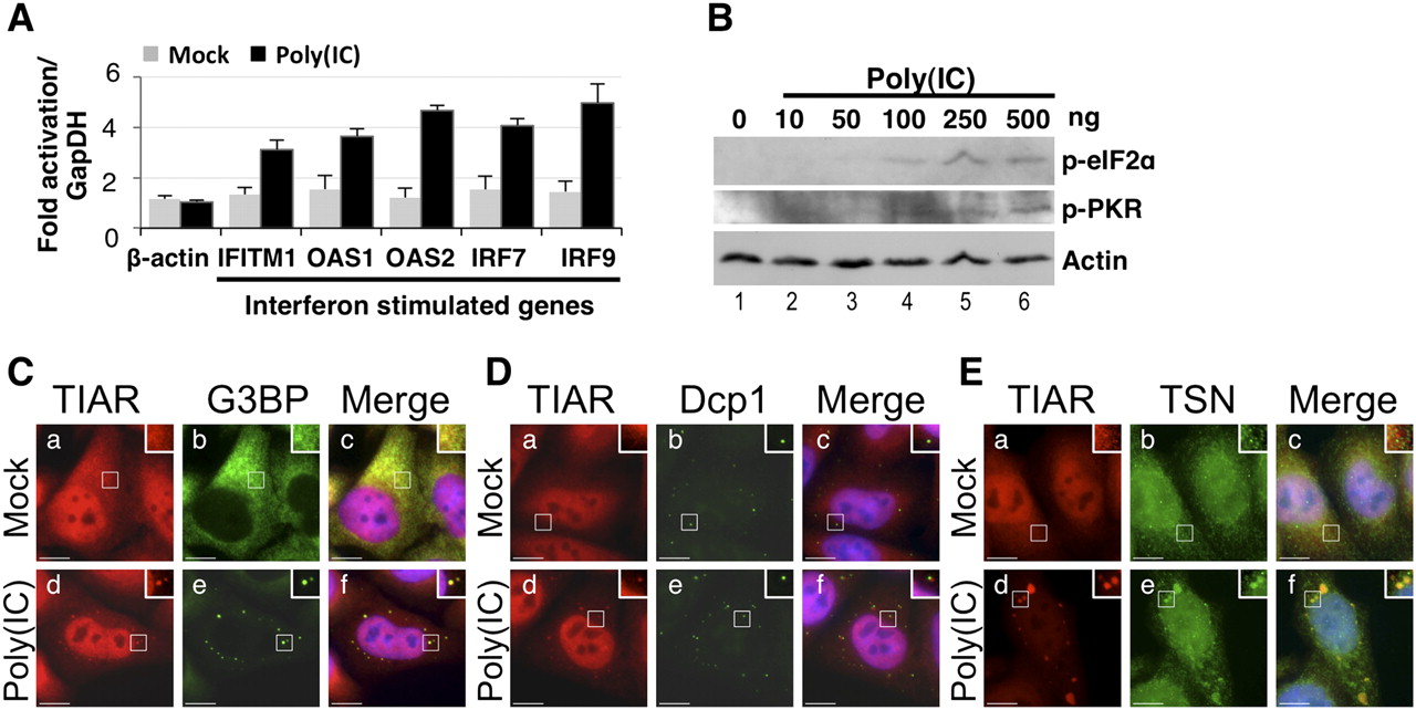
Stress granules are induced by poly(IC). (A) HeLa cells were mock-transfected or transfected with poly(IC), and RT/qPCR was used to quantify expression of β-actin or ISGs (IFITM1, OAS2, OAS1, IRF7, and IRF9) after 7 h. Fold-change in mRNA levels were calculated relative to those without poly(IC) (mock), and normalized to GapDH. Error bars are mean ± s.d. (n ≥ 3). (B) HeLa cells were transfected with 0–500 ng poly(IC), and cell lysates were prepared after 7 h. Immunoblotting was used to analyze phosphorylation of eIF2α (p-eIF2α) and PKR (p-PKR). Actin was a loading control. (C–E) HeLa cells were transfected with poly(IC), then fixed and processed after 7 h cells for immunofluorescence microscopy. DAPI staining is blue (Merge; c,f). (C) HeLa cells were stained for TIAR (red; a,d) and G3BP (green; b,e). (D) HeLa cells were stained for TIAR (red; a,d) and Dcp1 (green; b,e). (E) HeLa cells were stained for TIAR (red; a,d) and Tudor-SN (TSN) (green; b,e). Bar, 10 μm.




