


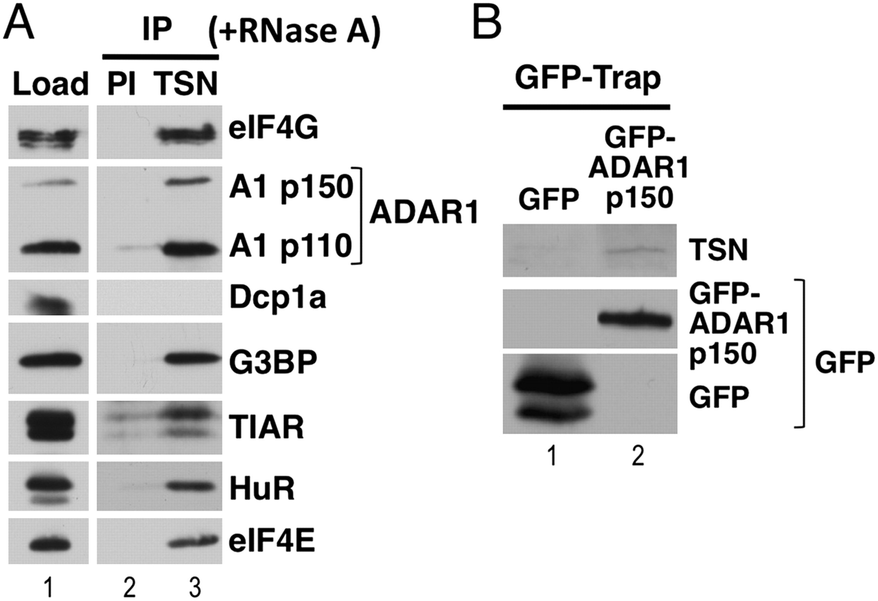
Tudor-SN interacts with stress granule components. (A) A Tudor-SN (TSN) antibody was used to immunoprecipitate proteins from RNase A-treated HeLa cell lysates, and immunoblotting was subsequently used to detect interacting proteins. Pre-immune serum (PI) was used as a control. Total protein (Load) is shown. (B) HeLa cells were transiently transfected with expression vectors for GFP (control) or GFP–ADAR1 p150, and lysates prepared after 24 h. GFP–Trap was subsequently used to immunoprecipitate GFP or GFP–ADAR1 p150, and immunoblotting used to analyze the immunoprecipitates. A specific Tudor-SN antibody (TSN) was used to detect Tudor-SN. The immunoblot was additionally probed with anti-GFP to verify expression of GFP and GFP–ADAR1 p150.




