


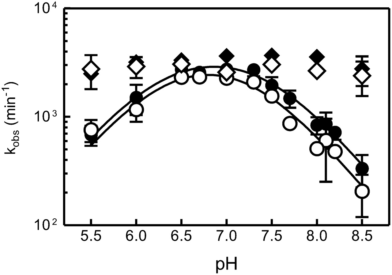
pH dependence of fluorescence-detected folding of the S. mansoni hammerhead ribozyme. The folding experiments at different pH values were performed in a stopped-flow fluorimeter by addition of Mg2+ (10 mM) to a pre-annealed complex of ribozyme and noncleavable substrate (1 μM). The folding reactions were carried out at 25°C in the presence of 100 mM NaCl and 50 mM MES (pH 5–6), MOPS (pH 6.5–7.5), HEPES (pH 7.0–8.0), TRIS (pH 7.5–8.5), TAPS (pH 8.2–8.9), and CHES (pH 8.5–9.0). The folding traces were fitted as described in Materials and Methods. Extracted, log apparent rate constants kobs (per minute) were plotted as a function of pH. The differences in the apparent rate of folding for loop pyC1.9 (black diamonds) and Stem I pyC1.1 (open diamonds) at different pH values did not exceed 10%. The apparent rate of folding for core residues pyC7 (black circles) and pyC3 (open circles) shows at least a one order of magnitude lower rate of folding at high and low pHs as compared with pH 7.0. The apparent bell-shaped curve could be fitted to a Henderson–Hasselbalch-type equation (see Materials and Methods) yielding two apparent pKa values of 6.3 (±0.2) and 7.4 (±0.1).




