


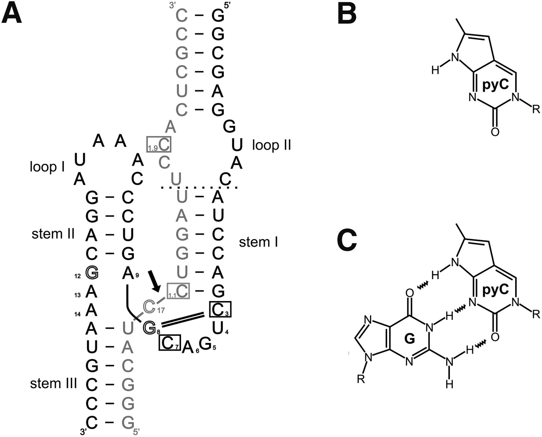
(A) Secondary structure of the Schistosoma mansoni hammerhead ribozyme construct used in this study. This representation emphasizes the global geometry as seen in the first crystal structure of the S. mansoni ribozyme (Martick and Scott 2006). Enzyme strand (black); the substrate (gray); and positions of nucleotides replaced with the fluorescent analog (boxed). Outlined letters (C17, G8, and G12) indicate core nucleotides essential for catalysis, and the cleavage site is marked with a black arrow; the canonical 8-3 Watson-Crick base pair is shown as a black double line. Thick black and gray lines indicate backbone continuity, where the sequence has been separated for diagrammatic clarity. Base numbering is according to Hertel (Hertel et al. 1992). The dotted line in the region of Loop 1 shows the truncation position of the substrate and ribozyme strands to form the minimal form of the ribozyme lacking the tertiary interaction. (B) Structure of pyrrolo-cytosine (pyC) and (C) pyrrolo-cytosine–guanosine base pair (pyC-G).




