


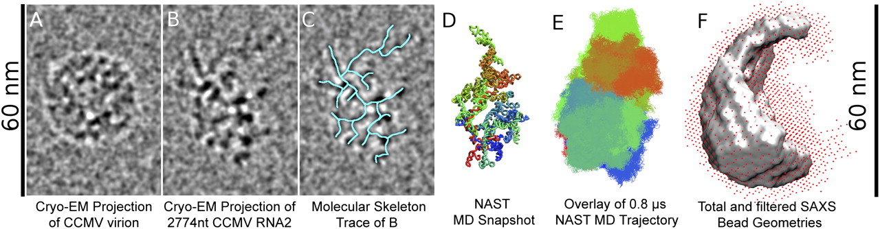
Comparison of cryo-EM, coarse-grained MD, and SAXS structures of 2777-nt CCMV RNA2 in physiological buffer with a wild-type virus particle. (Left) cryo-EM projections of a single wild-type CCMV virion (A) and one molecule of 2777-nt CCMV RNA2 (B) from a solution of RNA in an in vitro assembly buffer to which a trace amount of virions was added as a size marker. The parent image with projections of several RNA molecules is Figure 6B. Notice that the RNA molecule is comparable in size to the virion, but elongated and slightly larger in dimension. (C) A traced molecular skeleton (cyan) overlaid on the cryo-EM projection in panel B. (D) A NAST snapshot of the predicted minimum-free-energy secondary structure calculated from the primary sequence of CCMV RNA2. The RNA backbone is colored using a red–green–blue gradient from 5′ to 3′. Notice the striking similarities in shape and size between the molecular skeleton and the CGMD snapshot. (E) Two thousand snapshots from a 0.8-μsec MD trajectory are overlaid to illustrate the mobility of different parts of the molecule and the diversity of conformations in the MD ensemble. (F) The total volume (red dots) obtained by superimposing 50 SAXS bead reconstructions. The gray surface denotes a filtered consensus volume conserved in most bead reconstructions (see Materials and Methods), representing the most likely volume of the ensemble of molecules in solution. Notice the similarity in shape and size of the gray surface and the MD ensemble in panel E. Orthogonal views of the CGMD and SAXS structures and their shadow projections are compared with that of a model scalene ellipsoid in Supplemental Figure S7.




