


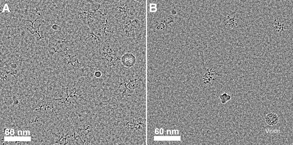
Cryo-EM projections of 2777-nt CCMV RNA2 in TE and in vitro assembly buffers. (A) Projections of randomly oriented RNA molecules in TE buffer, with a 30-nm spherical polystyrene (PS) bead used as internal size reference. (B) Four molecules of the same RNA in assembly buffer (see Materials and Methods) with a wild-type CCMV virus particle (28-nm outer diameter) added as an internal size marker. The central RNA molecule in panel B is shown in Figure 7 after rotation. Notice that in TE buffer the branching within each projected molecule is discernible and the individual arms appear to be longer and straighter (implying a longer persistence length). The molecules occupy smaller projected areas in the Mg2+-containing buffer consistent with their compaction and a reduction in the diversity of conformations. Traced skeletons for both panels are available as Supplemental Figure S6.




