


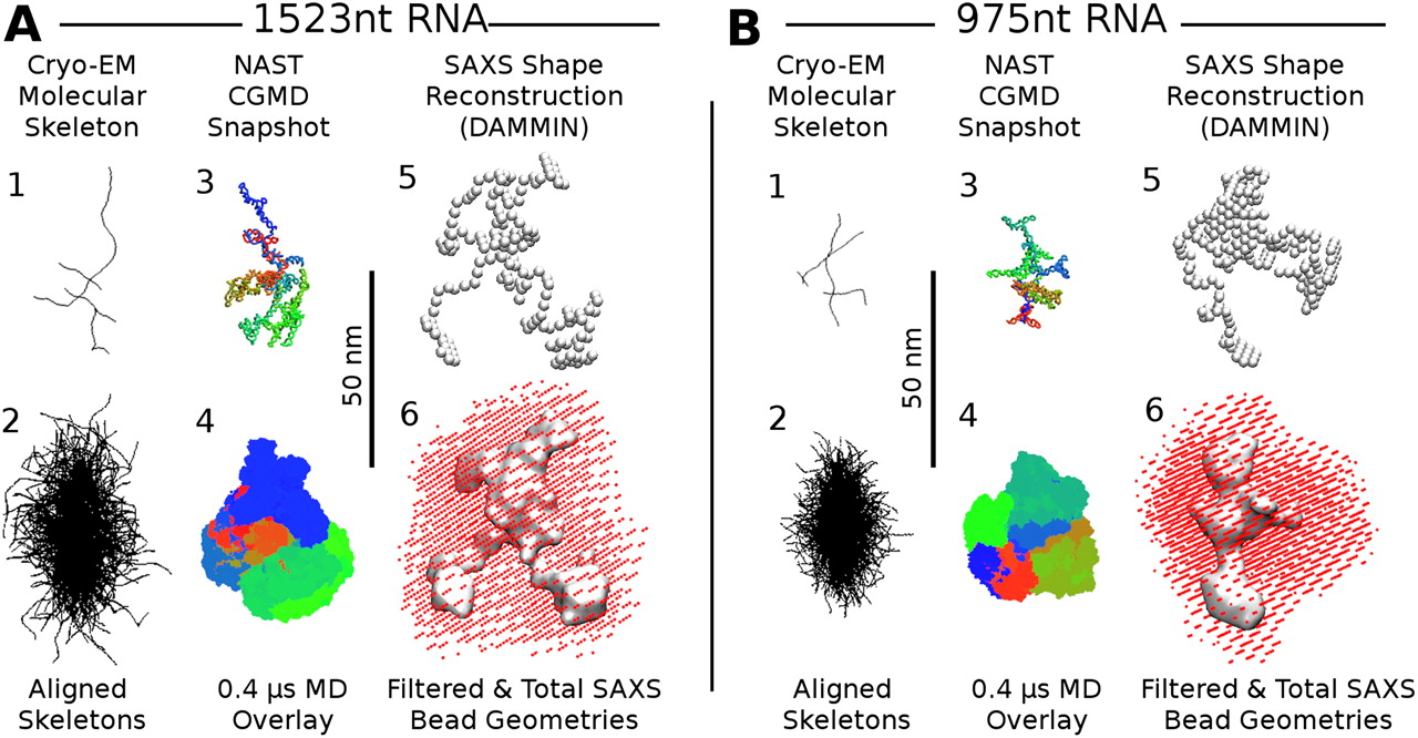
Comparison of structures from cryo-EM, coarse-grained MD, and SAXS, for the 1523-nt (A, left) and 975-nt (B, right) RNA molecules. The top rows in A and B show snapshots of (1) a traced single-molecule cryo-EM projection (TE buffer); (3) a NAST CGMD configuration showing the C3 backbone of the MFE secondary structure of the RNA, with a red–green–blue gradient indicating nucleotide index from 5′ to 3′; and (5) the centroid SAXS bead reconstruction. The bottom rows in A and B show composite images of ensembles of molecules: (2) 100 traced cryo-EM projections (TE buffer) with maximum Feret diameters vertically aligned with centers of mass in registry; (4) 1000 overlaid snapshots from a 0.4-μsec NAST trajectory aligned by minimizing RMSDs between corresponding nucleotides in each pair of snapshots by rotation; and (6) the ensemble volume of 50 overlaid SAXS bead reconstructions (red dots) and the filtered consensus volume (gray surface) (see text; Materials and Methods). The flatness of the molecules is revealed by comparing these composite images with the orthogonal views presented in Supplemental Figure S4.




