


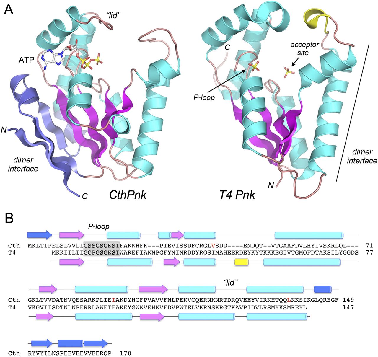
Tertiary structure of the CthPnkp kinase domain and comparison to phage T4 polynucleotide kinase. (A) The tertiary structures of the SeMet-kinase protomer A (CthPnk) and the kinase domain of bacteriophage T4 Pnkp (T4Pnk; from pdb 1LY1) were superimposed and then offset horizontally. Common secondary structural elements in CthPnk and T4Pnk are colored magenta (for β strands) and cyan (for α helices). Unique secondary structure elements in CthPnk that form the dimer interface are colored blue. N and C denote the N and C termini of the kinase polypeptides. ATP in the CthPnk active site is shown as a stick model. T4Pnk has two sulfate anions (shown as stick models) in the active site. The sulfate at left is in the NTP phosphate donor site and is coordinated by the P-loop, as indicated by the arrow. The 5′-OH acceptor site in the T4Pnk structure is demarcated by a second sulfate at right that mimics the 5′-terminal phosphodiester of the polynucleotide, HONpN–. (B) The secondary structure elements of CthPnk and T4Pnk (colored as in panel A) are shown above and below their aligned amino acid sequences, with β strands rendered as arrows and α helices as cylinders. Gaps in the alignment are indicated by dashes. The P-loop motifs are highlighted in gray boxes. Three native amino acid residues that were replaced by methionine in the SeMet-kinase are indicated in red.




