


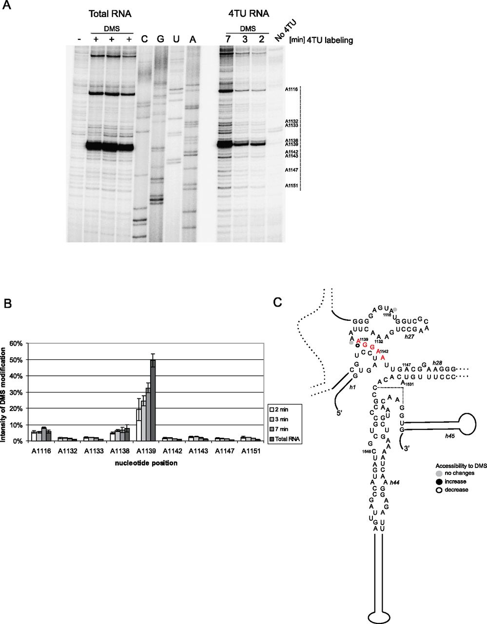
Changes in base-pairing in the central part of 18S rRNA revealed by different access to DMS modification in newly synthesized RNA. (A) DMS modification in vivo of the central part of 18S rRNA. The 4TU labeling was performed for 2, 3, and 7 min, with DMS added to the culture for the final minute. DMS modification and RNA purification were performed as described in the legend of Figure 3 and the text. Four sequencing lanes are presented. Primer 101 (complementary to 18S H31) was used for primer extension through the central pseudoknot region. The nucleotide positions are marked on the right side of the gel. (Dotted line along the autoradiogram) The region that was quantified. The three “Total RNA” lanes are technical replicates. (B) Semi-quantitative analysis of the DMS modification of total RNA relative to newly synthesized RNAs. The bars represent averages, with error bars showing standard deviations based on biological replicates from three independent experiments. (C) Changes in the intensity of DMS modifications in newly synthesized RNA compared with total RNA displayed on the secondary structure model of the analyzed part of rRNA. The modifications whose intensity is below 2.5% are not presented on the structure. (Red) rRNA sequence involved in interactions with the U3 snoRNA.




