


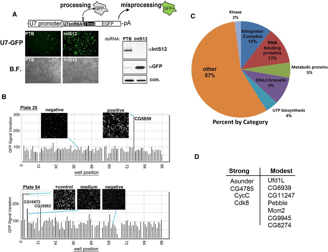
FIGURE 1.
Using the U7-GFP reporter to conduct a genome-wide RNAi screen. (A) Schematic of the U7-GFP reporter and the results from transient transfection of the reporter into S2 cells treated with dsRNA targeting PTB (−control) or IntS12 (+control). (Left) Fluorescence and bright-field images; (right) Western blot analysis. (B) Bar graph representing quantification of screen results for plates 25 and 54. Insets are taken from the acquired image collection. (C) Pie graph representing results of RNAi screen categorically. (D) Trimmed-down list of non-Integrator proteins that reproducibly scored as strong or modest by secondary screening.




